Download
1 / 53
641 likes | 3.56k Views
Head injury. Presented by: Remya Gopinath. DEMOGRAPHIC DATA. Name: Case No.4 MR No : 185840 Diagnosis : RTA WITH HEAD INJURY Age: 6 YRS Gender: Male Date of admission: 2/10/2012.

E N D
Head injury • Presented by: Remya Gopinath
DEMOGRAPHIC DATA Name: Case No.4 MR No : 185840 Diagnosis : RTA WITH HEAD INJURY Age: 6YRS Gender: Male Date of admission: 2/10/2012
PHYSICAL ASSESSMENT GENERAL ASSESSMENT: Patient is bedridden, lying over bed with tracheostomy and NGT in situ. SKIN: Normal in state, warm To touch. No sores or redness present all over the body. HEAD AND NECK: Head is slightly extended. No visible injury noted in the scalp area.Involuntary eye movement present.5mm tracheostomy tube present over neck region.
RESPIRATORY: Respiration through tracheostomy tube with in normal rate. Cough with mild to moderate secretion present.spo2 maintaining on room air. Thorax is symmetrical in size. CARDIOVASCULAR: No deformities noted. GENITOURINARY: self voiding on diaper GASTROINTESTINAL : Abdomen is soft, not distended. Feeding via NGT.Bowel sound present.
MUSCULO-SKELETAL : All limbs are spastic with flexed upper extremities and extended lower extremities. Mild spontaneous limb movement present. Displaced fracture is seen in middle of left clavicle. NEUROLOGY : Patient is semi conscious .pupils are bilaterally reacting to light.Bilatral flexure response to painful stimuli.GCS E4VTM3.
Patient history Past medical history Patient was in normal healthy living until the day of accident. Present medical history Patient received in ER on 2/10/2012 after being involved in RTA with an unconscious and irritable state. Vomiting and loc at scene for 5 minutes. On examination vital signs are Pulse-103/mt, BP-120/70,Temp,36.7 *c ,SPO2 -94%,GCS 8/15. Pupils are bilaterally reacting to light. Limb movements are equal and normal in all 4 limb.NCCT brain shows SAH in right fronto_parietallobe,diffuse brain edema,small hemorrhage in the 4th ventricle and opacification in all paranasal sinuses. Scalp swelling is seen in left parital area and no fracture is seen in cranial vault.
After the initial management patient shifted to ICU .On 8th day of admission ,patient developed tachypnoea,for which he investigated and found to have collapse of right lung. Intubation done and relaxant started with Inj.Midazolam and Inj.Fentanyl.Tracheostomy done on 13/12/2012.The repeat NCCT on 28/10/2012,which shows subdural haematoma with midline shift 4mm.Right frontal and parietal burrhole with evacuation of subdural hygroma done. After 2 months of admission clinically patient is opening eyes, bilateral flexure response to pain. All limbs are spastic,pupils both equal reacting ,afebrile,on NGT feeding with pediasure q 4h.patient shifted to pedia ward for further management.
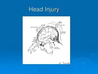
HEAD INJURY Definition It is an injury to the skull or brain that is severe enough to interfere with normal functioning.
Anatomy& Physiology The brain is one of the largest and most complex organs in our body. It controls our body, receives information, analysis information and stores information. It is made up of more than 100 billion nerves that communicates in trillions of connections called synapse. The skull consisting of 22 bones all together.These bones are divided into 8 cranial bones and 14 facial bones. Cranial bones form the cranial cavity and protects the brain.
There are typically 206 bones in the body.Out of these there are 22 bones of the Skull, which include: • 8 Cranial Bones: 1 x Ethmoid Bone1 x Frontal Bone1 x Occipital Bone2 x Parietal Bones1 x Sphenoid Bone 2 x Temporal Bones
14 Facial Bones: 2 x Inferior Nasal Conchae2 x Lacrimal Bones1 x Mandible2 x Maxillae (pl.); Maxilla (sing.)2 x Nasal Bones 2 x Palatine Bones1 x Vomer2 x Zygomatic Bones
CRANIAL NERVES Olfactory I: sense of smell. Optic Nerve II: sight of retina. Oculomotor Nerve III: eye movement and pupil constriction. Trochlear Nerve IV: eye movements. Trigeminal Nerve V: carry somatosensory information to face, head and chewing muscles of jaws. Abducens Nerve VI: eye movement. Facial VII: control the muscles used for facial expressions (smiling, frowning etc). It also stimulates salivary glands to produce saliva.
Vestibulocochlear VIII: hearing and balance. Glossopharyngeal IX: taste sensation ,gag reflexes. Vagus X: It carries somatosensory information from organs of thoracic, abdominal cavity including heart and from that of gastrointestinal tract. Spinal Accessory Nerve XI: leads to muscles of neck, back and larynx. It controls the head movement. Hypoglossal Nerve XII:controls the muscles of tongue
Meninges Meninges are the connective tissue membrane enclosing the brain and the spinal cord. It is divided into 3.outer most duramater,arachanoid mater and the inner most piamater.
Lobes of brain Frontal lobe: is responsible for problem solving,judgement and motor function. Parietal lobe: manage sensation, hand writing and body position. Temporal lobe: is involved with memory and hearing. Occipital lobe: contain the brains visual processing system.
Sutures of brain Coronal suture: present between frontal and parietal bones. Lambdoid suture: present between occipital and parietal bones. Sagital sutures: present between two parietal bones. Squamous sutures: present between parietal and temporal bones.
Major Regions of Brain Brain is divided into 3 major parts • Cerebrum • cerebellum • Brain stem
Cerebrum: Cerebrum is the most superior part of the brain. It is made up of by thick gray matter as surface layer and internally with white matter.It consist of thalamus, hypothalamus and epithalamus.
Cerebellum: Cerebellum located dorsal to the pons and medulla. It receives the impulses from cerebral motor cortex, various stem and sensory receptors in order to control skeletal muscle contraction.
Brain stem: Brain stem is similarly structured as the spinal cord. It is divided in to midbrain ,pons and medulla oblongata.mid brain acts as a fibre pathway between higher and lower brain centres.The pons mainly a conduction region also contribute to the regulation of respiration and cranial nerves. Medulla oblongata regulate the respiratory rhythm, heart rate,B P etc...
Blood supply to the brain The major arteries are the vertebral and internal carotid arteries. This communicating arteries forms the circle of willis,which equalizes the blood pressure in the brain’s anterior and posterior region.
Pathophysiology Damage to the brain from traumatic injury takes two forms Primary: Initial damage to the brain that result from the traumatic event. Secondary: It occurs hours and days after the initial injury and result from inadequate delivery of oxygen and nutrients.
Brain suffers traumatic injury Brain swelling or bleeding increase intracranial volume Increased ICP . Pressure on blood vessels causes blood flow to the brain to slow Cerebral hypoxia or ischemia Continues increase in ICP Brain herniation Cerebral blood flow cease Brain death
Types of head injury Concussion: Transient interruption in brain activity. No structural injury noted on radiographs Contusion: Bruising of the brain with associated swelling. Intra cranial haemorrhage: Bleeding in to the brain tissue commonly associated with edema.
Epidural hematoma: Blood between inner table of skull and dura.Associated with injury or laceration of the middle meningeal artery secondary to a temporal bone fracture. Subdural hematoma: Blood between the dura and arachnoid space caused by venous bleeding. Commonly associated with ICH or contusion. Diffuse axonal injury or shear injury: Axonal tear with in the white matter of the brain. Frequently occurs with the corpus callosum or brain stem and at the frontal or temporal poles associated with prolonged coma.
Diagnostic procedures Diagnostic procedures Diagnostic procedures Diagnostic procedure
NERVE CONDUCTION VELOCITY A nerve conduction study (NCS) is a medical diagnostic test commonly used to evaluate the function, especially the ability of electrical conduction, of the motor and sensory nerves of the human body.
ELECTRONYSTAGMOGRAPHY is a diagnostic test to record involuntary movements of the eye caused by a condition known as nystagmus. It can also be used to diagnose the cause of vertigo, dizziness or balance dysfunction by testing the vestibular system.
Management All therapy is directs towards preserving brain homeostasis and preventing secondary brain injury. Treatment to prevent secondary injury includes stabilization of cardiovascular and respiratory function to maintain adequate cerebral perfusion, control of haemorrhage,hypovolemia and maintaining of blood gas values.
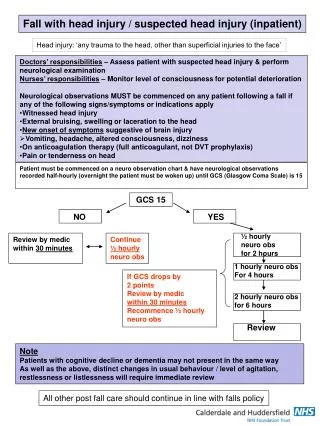
Nursing assessment Assessment • Collection of history • GCS score • Neurologic status • Presence of CSF leakage • Pupillary response to light
Severe head injury ATLS Evaluation Initial management Intubation with ventilation and sedation Fluid resuscitation CT Brain OT Surgical lesion YES NO MONITOR ICP ICU Treat intra cranial hypertension
Complications Infection-respiratory Hydrocephalus Post traumatic seizure Permanent neurologic deficit Coma Chronic headache Death