Download
1 / 52
520 likes | 526 Views
CYTOKINES , SURFACE MOLECULES, ANTIBODIES. Jan Novák. The Immune System is the Third Line of Defense Against Infection. Clonal Selection of B Cells is Caused by Antigenic Stimulation. Humoral Immunity - apoptosis Programmed cell death (“Falling away”).

E N D
CYTOKINES, SURFACE MOLECULES, ANTIBODIES Jan Novák
The Immune System is the Third Line of Defense Against Infection
Clonal Selection of B Cells is Caused by Antigenic Stimulation
Humoral Immunity - apoptosis • Programmed cell death (“Falling away”). • Human body makes 100 million lymphocytes every day. If an equivalent number doesn’t die, will develop leukemia. • B cells that do not encounter stimulating antigen will self-destruct and send signals to phagocytes to dispose of their remains. • Many virus infected cells will undergo apoptosis, to help prevent spread of the infection.
Immunological Memory • Antibody Titer: The amount of antibody in the serum. • Pattern of Antibody Levels During Infection • Primary Response: • After initial exposure to antigen, no antibodies are found in serum for several days. • A gradual increase in titer, first of IgM and then of IgG is observed. • Most B cells become plasma cells, but some B cells become long living memory cells. • Gradual decline of antibodies follows.
Antibody Structure • Monomer: A flexible Y-shaped molecule with four protein chains: • 2 identical light chains • 2 identical heavy chains • Variable Regions: Two sections at the end of Y’s arms. Contain the antigen binding sites (Fab). Identical on the same antibody, but vary from one antibody to another. • Constant Regions: Stem of monomer and lower parts of Y arms. • Fc region: Stem of monomer only. Important because they can bind to complement or cells.
Consequences of Antigen-Antibody Binding • 1. Agglutination: Antibodies cause antigens (microbes) to clump together. • IgM (decavalent) is more effective that IgG (bivalent). • Hemagglutination: Agglutination of red blood cells. Used to determine ABO blood types and to detect influenza and measles viruses. • 2. Opsonization: Antigen (microbe) is covered with antibodies that enhances its ingestion and lysis by phagocytic cells.
Humoral Immunity • 3. Neutralization: IgG inactivates viruses by binding to their surface and neutralize toxins by blocking their active sites. • 4. Antibody-dependent cell-mediated cytotoxicity: Used to destroy large organisms (e.g.: worms). Target organism is coated with antibodies and bombarded with chemicals from nonspecific immune cells. • 5. Complement Activation: Both IgG and IgM trigger the complement system which results in cell lysis and inflammation.
Immunoglobulin Classes • I. IgG • Structure: Monomer • Percentage serum antibodies: 80% • Location: Blood, lymph, intestine • Half-life in serum: 23 days • Complement Fixation: Yes • Placental Transfer: Yes • Known Functions: Enhances phagocytosis, neutralizes toxins and viruses, protects fetus and newborn.
Immunoglobulin Classes • II. IgM • Structure: Pentamer • Percentage serum antibodies: 5-10% • Location: Blood, lymph, B cell surface (monomer) • Half-life in serum: 5 days • Complement Fixation: Yes • Placental Transfer: No • Known Functions: First antibodies produced during an infection. Effective against microbes and agglutinating antigens.
Immunoglobulin Classes • III. IgA • Structure: Dimer • Percentage serum antibodies: 10-15% • Location: Secretions (tears, saliva, intestine, milk), blood and lymph. • Half-life in serum: 6 days • Complement Fixation: No • Placental Transfer: No • Known Functions: Localized protection of mucosal surfaces. Provides immunity to infant digestive tract.
Immunoglobulin Classes • IV. IgD • Structure: Monomer • Percentage serum antibodies: 0.2% • Location: B-cell surface, blood, and lymph • Half-life in serum: 3 days • Complement Fixation: No • Placental Transfer: No • Known Functions: In serum function is unknown. On B cell surface, initiate immune response.
Immunoglobulin Classes • V. IgE • Structure: Monomer • Percentage serum antibodies: 0.002% • Location: Bound to mast cells and basophils throughout body. Blood. • Half-life in serum: 2 days • Complement Fixation: No • Placental Transfer: No • Known Functions: Allergic reactions. Possibly lysis of worms.
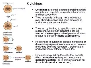
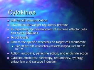
CYTOKINES a diverse group of non-antibody proteins released by cells that act as intercellular mediators, especially in immune processes
CYTOKINES Low molecular weight soluble proteins (polypeptides) produced in response to microbes and other antigens They act via cell surface receptors to mediate and regulate the amplitude and duration of the immune-inflammatory responses, through activation of macrophages, controlling growth and differentiation of T and B cells
Naming of Cytokines • 1. Monokines - produced by mononuclear phagocytes (monocytes) • 2. Lymphokines - produced by activated T cells, primarily helper T cells • 3. Interleukins - cytokines made by one leukocyte and acting on other leukocytes 4.Chemokines-cytokines with chemotactic activities
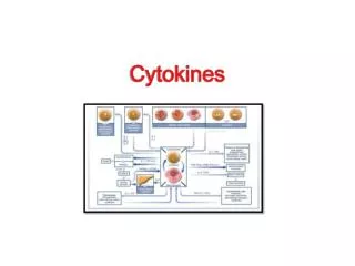
Classification of Cytokines • Interleukins - cytokines made by one leukocyte and acting on other leukocytes • Chemokines- cytokines with chemotactic activities • Interferons – tissue antiviral cytokines • Transforming growth factors – growth, regulation • Colony stimulating factors – growth, stimulation • Tumor necrosis factors – induction of apoptosis
Properties • 1.Produced by cells involved in both natural and specific immunity • 2. Mediate and regulate immune and inflammatory responses • 3. Secretion is brief and limited not stored as pre-formed molecules. Synthesis is initiated by new short-lived gene transcription mRNA is short-lived
5. Redundancy-similar functions can be stimulated by different cytokines. Receptors for cytokines are heterodimers (sometimes heterotrimers) that can be grouped into families in which one subunit is common to all members of a given family. (hard clinical diagnostics)
Properties • 6. Often influence the synthesis of other cytokines. They can produce cascades, or enhance orsuppress production of other cytokines. They exert positive or negative regulatory mechanisms for immune inflammatory responses • 7. Often influence the action of other cytokines. • antagonistic -cytokines causing opposing activities • additive • synergistic -two or more cytokines acting together (greater than additive)
Properties • 8. Bind to specific receptors on target cells with high affinity.. • 9. Cellular responses to cytokines are generally slow (hours), require new mRNA and protein synthesis
Functional Categories of Cytokines 1) Proinflammatory cytokines - Produced by activated microphages and NK cells in response to microbial infection - chemotaxis - killing - IL1, IL-6, IL-8, IL-12, IL-18, TNF-a
Functional Categories of Cytokines 2) Anti-inflammatory cytokines - Produced mainly by T cells - IL1Ra, IL-4, IL-10, TGF-b
Functional Categories of Cytokines 3) Growth factors - IL2, IL-3, IL-4, IL-5, IL-6, IL-7, IL-9, IL-11, IL-14, IL-15, G-CSF, EPO…
Functional Categories of Cytokines 4) Cytokines of humoral immunity - IL-4, IL-5, IL-9, IL-13
Functional Categories of Cytokines 5) Cytokines of cellular immunity - IL-2, IL-12, IFN-g
Functional Categories of Cytokines 6) Cytokines with antiviral potential - IFN-a,b,g,
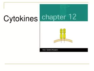
Cytokine Receptors • Divided into several families based on their structure and activities
Hematopoietin family • receptors are dimers or trimers • conserved cysteines in their extracellular domains and a conserved Trp-Ser-X-Trp-Ser sequence. • Examples are receptors for IL-2 through IL-7 and GM-CSF.
Interferon family • receptors have the conserved cysteine residues but not the Trp-Ser-X-Trp-Ser sequence, • Eg. the receptors for IFNa, IFNb, and IFNg.
Tumor Necrosis Factor family • receptors have four extracellular domains; they include receptors for soluble TNFa and TNFb as well as membrane-bound CD40 (important for B cell and macrophage activation) and Fas (which signals the cell to undergo apoptosis).
Chemokine family • receptors have seven transmembrane helices and interact with G protein. This family includes receptors for IL-8, MIP-1 and RANTES. • Chemokine receptors CCR5 and CXCR4 are used by HIV to preferentially enter either macrophages or T cells.
Therapeutic Uses of Cytokines 1) Interferon in treatment of viral diseases, cancer 2) Several cytokines are used to enhance T-cell activation in immunofideficincy diseases, e.g. IL-2, IFN-,TNF- 3) IL-2 and lymphokine activating killer cells (LAK) in treatment of cancer 4) GM-CSF induces increase in white cell count, it is used: a- To restore leukocytic count after cytotoxic chemotherapy induced neutropenia b- After bon marrow transplantation C- To correct AIDS-associated leukopenia
5) Anti-cytokines antibodies in management of autoimmune diseases and transplant rejection: a- Anti-TNF in treatment rheumatoid arthritis b- Anti-IL2R to reduce graft rejection c- Anti-TNF antibodies in treating septic shock d- Anti-IL-2R in treating adult T-cell leukemia e- Anti-IL-4 is under trial for treatment of allergies
Adhesive molecules • Adhesion of cells • Signal transduction
Groups of adhesive molecules • Integrins • Structures of intercellular matrix • Formed by two subunites (a and b) • Quiscent and activated conformation • LFA-1, VLA-1-5, CR3, CR4
Groups of adhesive molecules • Molecules of immunoglobulin structure • ICAM, CD80/86, CD2
Groups of adhesive molecules • Selectins and Lectins • L (leucotytes) • E (endothelial) • P (platelet) • Interactions between leukocytes and endothelium • Receptors of NK cells (Lektins of C type)
Groups of adhesive molecules • Mucins • CD43 • Supression of interlekcocyte contact
FC receptors • Bind FC parts of immunoglobulins • Divided according to the class of antibody they bind • CD16, CD32, CD64
Complement receptors • Phagocytosis of opsonized particles (CR3,4) • Clearance of immunocomplexes • Receptors for chemotaxis (C3aR, C5aR)