Download
1 / 30
350 likes | 920 Views
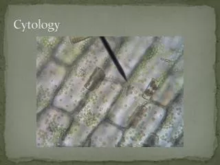
Cytology of Body Fluid. Edmund S, Cytology, Chapter 4: Pleural, pericardial, and peritoneal fluid Richard M DeMay, The art & science of cytopathology, Chapter 8: Fluid Leopold G.Koss, Koss’ diagnostic cytology, Chapter 26: Effusion in the presence of cancer. Speaker : 黃筱琪

E N D
Cytology of Body Fluid Edmund S, Cytology, Chapter 4: Pleural, pericardial, and peritoneal fluid Richard M DeMay, The art & science of cytopathology, Chapter 8: Fluid Leopold G.Koss, Koss’ diagnostic cytology, Chapter 26: Effusion in the presence of cancer Speaker : 黃筱琪 Advisor : 聶鑫 主任 李修南 學姊 Date : 3.15.2006
Outline • Representation of the three body cavities • Collection and preparation of specimen • Benign elements • Non-neoplastic conditions • Malignant effusions---primary tumors ---metastatic tumors • Differences Between Adenocarcinoma and Mesothelioma
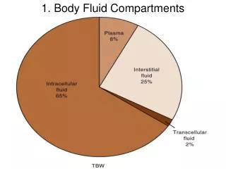
Transudates Exudate • Increased hydrostatic pressure: Congestive heart failure • Decreased oncotic pressure: cirrhosis, nephrosis, and malnutrition • (decreased albumin) • Inflammation: Infection, infarction, hemorrhage • Tumor Accumulation of fluids in body cavities
Cytocentrifuge preparation Cell block Heparinized bottles (3 units heparin/ml) Unfixed Alcohol-fixed Adding plasma and thrombin solution Papanicolaou-stained Wrapped in filter paper Air-dried cytocentrifuge preparation Placed in a cassette Diff-Quik (Hematologic malignancy is suspected) Embedded in paraffin Cut and H&E stain Collection and preparation of specimen
Mesothelial cells • Usually dispersed as isolated cells • Binucleation and multinucleation • Occasional small clusters with “windows” • Dense cytoplasm with clear outer rim (lacy skirt) Benign elements Mesothelial cells
Reactive mesothelial cell • Pleomorphic and enlarged nuclei • Hyperchromasia • Prominent nucleoli • Mitotic figures
Histiocytes Other blood cells • Nuclei often kidney shape • Cytoplasm granular and vacuolated • No window between cells • CD68 positive • Lymphocytes • Eosinophils • Neutrophils • Plasma cells • Red blood cells
Acute serositis Eosinophilic effusions • Bacterial infection: pleural empyema, bacterial peritoneal • Color of the fluid: creamy pale yellow (purulent) • Cytology preparation: high cellular and polymorphonuclear leukocytes • Thoracic trauma, pneumothorax, hemothorax, pulmonary infarcts • Cytology preparation: high number of eosinophils • Eosinophilic pleural effusions more common • Charcot-Leyden crystals Non-neoplastic conditions Eosinophilic pleural fluid
Tuberculous pleuritis Rheumatoid pleuritis • Color of the fluid: turbid and greenish-yellow • Cytology preparation: high cellular of lymphocytes (T cells) • Differential diagnosis: inflammatory effusion of non-tuberculous origin • Necrotizing granulomatous inflammation (joint disease) • Cytology preparation: clumps of granular debris • multinucleated macrophages Tuberculous pleuritis Multinucleated macrophages
Systemic lupus erythematosus • Cytology preparation: Lupus erythematosus cell (LE cell) LE cell
Malignant mesothelioma • Clinical history: asbestos exposure, persistent pleural effusions, chest pain • Epithelial (carcinomatous) pattern Malignant effusions---primary tumors
Cell-in-cell pattern “More and bigger cells, in more and bigger clusters”
Adenocarcinoma • Large nucleoli • Secretory vacuoles • Three dimensional aggregates • Increased N/C ratio • Irregular nuclear membranes
Breast cancer Lung cancer • Cannonballs: • Tight packed large balls of cells • Smooth borders • Indian files
Ovarian carcinoma • Irregular clusters of cells • Large and clear vacuoles Gastric carcinoma • Signet ring cell pattern
Clear cell carcinoma of kidney cancer • Clear or granular and • vacuolated cytoplasm Papillary carcinoma of the thyroid • Psammoma bodies
Squamous cell carcinoma Small cell carcinoma • Keratinized or non-keratinized • Tadpoles and bizarre shape • Isolated and molded cells • Scant cytoplasm, inconspicuous nucleoli F4.27 F4.28
Non-Hodginkin lymphoma • Large cell lymphoma • Nuclei large than histiocyte • Eccentric nuclei • Abundant blue cytoplasm • Best appreciated in Diff-Quik • Follicular lymphoma • Irregular nuclear contours • Scant cytoplasm
Small lymphocytic lymphoma • Differential diagnosis: • chronic inflammation (tuberculosis) • lymphoblastic lymphoma • Small to medium sized lymphocytes • Fine powdery chromatin • Scant cytoplasm
Hodgkin lymphoma Multiple myeloma • Reed-Sternbery cells: • Multinucleated cell with • huge inclusion-like nucleoli • Single, lack cohesive aggregate • Numerous malignant plasma cells • Immunocytochemistry stain: • kappa and lambda light chain (+) • CD138 (+)
Melanoma Sarcomas • Isolated round cells with prominent nucleoli • Fine brown cytoplasmic pigmentation • Intranuclear pseudoinclusions • Immunocytochemistry stain: S-100(+), HMB-45(+) • Isolated cells Pleomorphic sarcoma Osteosarcoma Liposarcoma Large and bizarre shaped Round cell sarcoma Rhabdomyosarcoma Neuroblastoma Small and uniform shaped Spindle cell sarcoma Fibrosarcoma Leiomyosarcoma Spindle shaped
- + - + + - + - + - - + + - + - + + - + - + - + C: cytoplasm; M: membrane; N: nuclear
THE END Thank you for your attention