Download

1 / 19
210 likes | 404 Views
The Autonomic Nervous System. Dr. Nimir Dr. Safa. Objectives Review the subdivisions of the nervous system. Review the general arrangement and compare the sympathetic and parasympathetic parts. Describe the following plans Para vertebral ganglia. Prevertebral ganglia.

E N D
The Autonomic Nervous System Dr. Nimir Dr. Safa
Objectives • Review the subdivisions of the nervous system. • Review the general arrangement and compare the sympathetic and parasympathetic parts. • Describe the following plans • Para vertebral ganglia. • Prevertebral ganglia. • Parasympathetic ganglia. • Splanchnic nerves. • Autonomic plexuses • Map out the various plexuses in head and neck, thorax, abdomen and pelvis. • Make a list of the components of the system. • Review the basic structure of sympathetic trunk. • Describe the source of sympathetic system in the neck and make a list of target organs.
Describe the Para vertebral sympathetic ganglia in the abdomen, their locations and target organs. • Discuss the relation of this system to the adrenal medulla. • Discuss the sympathetic innervation of blood vessels. • Make a list of the components of the system. • Make a list of cranial nerves having parasympathetic activity. • Describe the parasympathetic ganglia in the head and neck, their locations and target organs. • Describe the sacral parasympathetic out flow. • Make a list of its target organs.
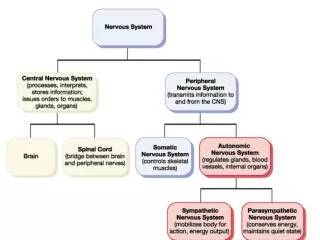
The nervous system is divided into : • Central nervous system,which consists of the brain and spinal cord. • Peripheral nervous system , which consists of the cranial and spinal nerves and their associated ganglia. • The autonomic nervous system is part of nervous system innervating involuntary structures, such as the heart, smooth muscle, and glands. • It isdivided into sympathetic and parasympathetic, with afferent and efferent nerve fibers.
The autonomic nervous system, like the somatic nervous system, has afferent, connector, and efferent neurons. • The afferent impulses originate in visceral receptors and travel via afferent pathways to the central nervous system. • They are integrated through connector neurons at different levels, then leave via efferent pathways to visceral effector organs.
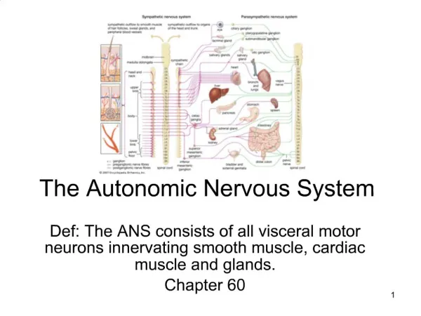
Sympathetic Part of the Autonomic System: • The sympathetic is larger and is widely distributed throughout the body, innervating heart, lungs, muscle in many blood vessels, hair follicles, sweat glands, and abdominopelvic viscera. • The sympathetic system consists of the efferent outflow from the spinal cord, two ganglionatedsympathetic trunks, important branches,plexuses, and regional ganglia.
Efferent Nerve Fibers (Sympathetic Outflow) • The lateral gray columns (horns) of the spinal cord(T1-L2)which contain cell bodies of the sympathetic connector neurons. • Myelinated axons (preganglionic)leave the cord in anterior roots and pass via white ramicommunicantes to the paravertebral ganglia of the sympathetic trunk.
Preganglionic fibers are distributed as follows: • 1. Synapse inparavertebral ganglia. • The postganglionic nonmyelinated axons pass to spinal nerves as gray rami communicantes. • They are distributed in branches of the spinal nerves to smooth muscle in the blood vessel walls(vasomotor), sweat glands(sudomotor), and arrector muscles of the hairs of the skin(pilomotor).
2. They go up to synapse in cervical ganglia and down to synapse in lumbar and sacral ganglia. • Postganglionic fibers join cervical(head & neck), lumbar, sacral, and coccygeal spinal nerves .
3. Pass through the ganglia of the sympathetic trunk without synapsing. These myelinated fibers leave the sympathetic trunk as: • Greater splanchnicwhich synapse in celiac plexus, renal plexus, and suprarenal medulla. • Lesser splanchnicwhich synapse in lower part of the celiac plexus. • Lowest or least splanchnicnerves which synapse in renal plexus..
The splanchnic nerves, therefore, are formed of preganglionic fibers. • The postganglionic fibers are distributed to the smooth muscle and glands of the viscera. • The ratio of preganglionic to postganglionic sympathetic fibers is about 1:10, permitting a wide control of involuntary structures
Afferent Nerve Fibers • Afferent myelinatedfibers travel from viscera through the sympathetic ganglia without synapsing. • They pass to spinal nerves via white ramicommunicantes and reach their cell bodies in the posterior root ganglion of the corresponding spinal nerve. • The central axons then enter spinal cord and may form afferent component of a local reflex arc or ascend to higher centers, such as the hypothalamus.
Parasympathetic Part of the Autonomic System: • The activities of the parasympathetic part of the autonomic system are directed toward conserving and restoring energy. The heart rate is slowed, pupils are constricted, peristalsis and glandular activity is increased, sphincters are opened, and the bladder wall is contracted. • Efferent Nerve Fibers (Craniosacral Outflow) • The connector nerve cells of the parasympathetic part of the autonomic nervous system are located in the brainstem and the sacral segments of the spinal cord.
Those nerve cells located in the brainstem form nuclei in the following cranial nerves: • Oculomotor(parasympathetic or Edinger-Westphalnucleus). • Facial (superior salivatorynucleus and lacrimatorynucleus). • Glossopharyngeal(inferior salivatorynucleus). • Vagusnerves (dorsal nucleus of the vagus). • Axons of these connector nerve cells are myelinated and emerge from the brain within the cranial nerves. • The cranial parasympathetic ganglia are : • Ciliary, pterygopalatine, submandibular, and otic.
Sacral connector nerve cells are found in the gray matter of S2-S4. • These cells are not sufficiently numerous to form a lateral gray horn, as do the sympathetic connector neurons in the thoracolumbar region. • The myelinated axons leave the spinal cord in the anterior nerve roots of the corresponding spinal nerves. • They then leave the sacral nerves and form the pelvic splanchnic nerves .
The myelinated efferent fibers of the craniosacral outflow are preganglionic and synapse in: • Ganglia located close to the viscera they innervate. • Cranial parasympathetic ganglia which are ciliary(eye), pterygopalatine(nose), submandibular, and otic(salivary glands). • Plexuses, such as the cardiac , pulmonary, myenteric(Auerbach ), mucosal plexus (Meissner). • Myenteric & mucosal plexuses form enteric nervous system. • Pelvic splanchnicnerves synapse in ganglia in the hypogastric plexuses. Postganglionic parasympathetic fibers are short.