Download
1 / 33
350 likes | 536 Views
Measuring Action Potential Conduction Velocity and Determining the Site and Extent of Spinal Cord Injuries based on Sensory Deficits. Neuromuscular Junctions (NMJs) and Synaptic Delay. Action potentials in efferent axons arrive at NMJ

E N D
Measuring Action Potential Conduction VelocityandDetermining the Site and Extent of Spinal Cord Injuries based on Sensory Deficits
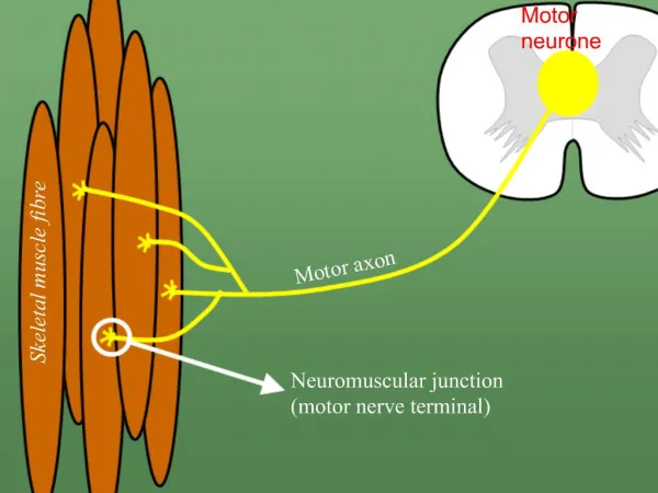
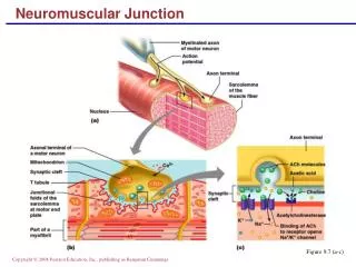
Neuromuscular Junctions (NMJs) and Synaptic Delay Action potentials in efferent axons arrive at NMJ Release of neurotransmitter (Acetylcholine)Depolarization of muscle cell membraneAppearance of action potential in muscle cellsMuscle contraction
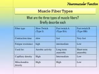
Choir = a muscle consisting of individual muscle cells (each singer) Choir output = sum of individual voices Electromyogram = sum of action potentials for all active muscle cells Single stimulus (command from conductor) produces Compound Muscle Action Potential
Peripheral nerve with afferent and efferent axons The Neuromuscular Junction andSynaptic Delay Cars = action potentials conducted along axons (lanes) Distance1/time1 Distance2/time2 Distance1-Distance2-/time1-time2 = highway speed = conduction velocity in axons!
An excellent resource on discriminating between the various causes of muscle weakness demonstrating the diagnostic power of transcutaneous electrical stimulation and recording compound muscle action potentials. Website Remember to measure the two distances!
PowerLab Scope 4.1 Settings: Input A = Ch 3, Input B = Off, Timebase = 50 ms, Samples = 2560 (40KHz) BioAmp range 10 mV or 5 mV or as necessary to see Compound Muscle Action PotentialDisplay: Overlay stimulator to Input A, Set up: Sampling = Sweep= Superimpose, Source= User, 0.5 sec delay, Display/Overlay All. Setup Stimulator: check Isolated, mode = pulse, delay = 10 ms, duration = 100 us, Amplitude = 20 mA. Measure latency from stimulus to onset (or first peak) of Compound Muscle Action Potential from Elbow and then from Wrist.
Which arm to test is determined randomly by last digit of SSN: • Even number test Right Arm • Odd number test Left Arm. Enter your data into the Spreadsheet on the Front Desk Computer
Role of Neurophysiologic Evaluation in Diagnosis Journal of the American Academy of Orthopaedic Surgeons May/June 200 Vol 8 No. 3 p 190-199 Lawrence R. Robinson, MD Dr. Robinson is Professor of Rehabilitation Medicine, University of Washington School of Medicine, Seattle, and Chief of Rehabilitation Medicine and Director, Electrodiagnostic Medicine Laboratory, Harborview Medical Center, Seattle. Reprint requests: Dr. Robinson, Rehabilitation Medicine, Harborview Medical Center, Box 359740, 325 Ninth Avenue, Seattle, WA 98104. Abstract The electrodiagnostic evaluation assesses the integrity of the lower-motor-neuron unit (i.e., peripheral nerves, neuromuscular junction, and muscle). Sensory- and motor-nerve conduction studies measure compound action potentials from nerve or muscle and are useful for assessing possible axon loss and/or demyelination. Needle electromyography measures electrical activity directly from muscle and provides information about the integrity of the motor unit; it can be used to detect loss of axons (denervation) as well as reinnervation. The electrodiagnostic examination is a useful tool for first detecting abnormalities and then distinguishing problems that affect the peripheral nervous system. In evaluating the patient with extremity trauma, it can differentiate neurapraxia from axonal transection and can be helpful in following the clinical course. In patients with complex physical findings, it is a useful adjunct that can help discriminate motor neuron disease from polyneuropathy or myeloradiculopathy due to spondylosis. Link to the abstract.
Left side of brain Right side of body Left side of body
Body-sense sensations • Proprioceptorsare receptors that give information about body position. • These receptors are located in muscles, tendons, ligaments, joints and skin. • Somesthetic sensations (senses associated with the surface of the body). • Mechanoreceptors detect pressure, force and vibration. These include: • Merkel's disks and Meissner's corpuscles in the superficial layer of the skin and, hair follicle receptors, Pacinian corpuscles and Ruffini's endings in deeper layers. • Thermoreceptors respond to temperature of receptor endings themselves. • Warm receptors respond to temperature between 30oC and 45o C with action potentials increasing as temperature increases. • Cold receptors respond to temperatures between 35o C and 20o C with action potentials increasing as the temperature falls. Both warm and cold receptors respond rapidly to temperature changes and show rapid adaptation. The brain uses the relative changes in the responses of hot and cold receptors to interpret the temperature of the environment. • Nociceptorstransduce harmful stimuli that we perceive as pain. These consist of free nerve endings. There are three types of nociceptors: • Mechanical - respond to intense mechanical stimuli. • Thermal - respond to intense heat. • Polymodal - respond to a variety of stimuli including mechanical, intense heat and chemicals released from damaged tissue.