Download
1 / 20
210 likes | 414 Views
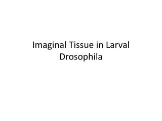
Synapses in Drosophila larval neuromuscular junctions. Drosophila NMJs.

E N D
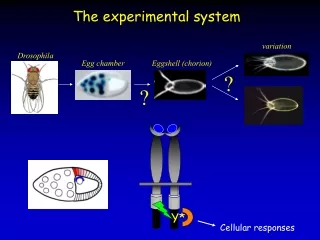
Drosophila NMJs In the developing NMJ of the Drosophila larva, a synapse that can be readily accessed for electrophysiology and easily imaged in a semi-intact preparation. The Drosophila larval NMJ shares important structural and molecular properties with mammalian CNS synapses. It is glutamatergic, with non-NMDA-type ionotropic glutamate receptors, and both the presynaptic active zones and postsynaptic specializations are organized through PDZ interactions in a manner similar to mammalian synapses. In addition, as in certain CNS synapses such as hippocampal CA1 inputs, the NMJ involves multiple neuronal connections onto the postsynaptic muscle. The NMJ also exhibits structural and functional plasticity. During larval development, muscle size increases more than 100-fold, causing a decrease in input resistance. To effectively depolarize and contract the muscle, synaptic currents must also increase as the larva grows. Two general mechanisms upregulate synaptic currents during development: one that regulates presynaptic structure and another that regulates transmission strength. During larval development, the degree of axonal branching as well as the number of boutons and active zones increase. Nascent boutons emerge either de novo or by budding from pre-existing boutons and come equipped with vesicles and active zones.
Drosophila neuromuscular junctions Muscle 6 Muscle 7
Synaptic structure at third instar NMJs Green: muscle maker (phalloidin) Muscle 6/7 synaptic boutons Red: synapse maker (Dlg)
Presynaptogenesis at fly NMJ B A D C
Retrograde BMP signaling regulates synaptic growth Green: MHC-CD8-GFP-SH (synaptic bouton maker)