Download
1 / 47
720 likes | 2.17k Views
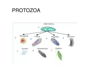
PROTOZOA. Presented By: Dr. Shaymaa Abdalal Medical Parasitology Demonstrator . INTESTINAL PROTOZOA. INTESTINAL PROTOZOA. Numerous protozoa inhabit the gastro-intestinal tract of humans .

E N D
PROTOZOA Presented By: Dr. Shaymaa Abdalal Medical Parasitology Demonstrator
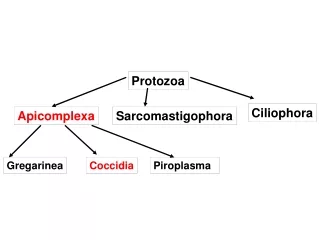
INTESTINAL PROTOZOA • Numerous protozoa inhabit the gastro-intestinal tract of humans . • Entamoebahistolytica can become a highly virulent and invasive organism that causes a potentially lethal systemic disease. • Giardialamblia can cause severe acute diarrhea which may lead to a chronic diarrhea and nutritional disorders
INTESTINAL PROTOZOA • Intestinal protozoa are transmitted by the fecal-oral route . • exhibit similar life cycles consisting of a cyst stage and a trophozoite stage
INTESTINAL PROTOZOA www.tulane.edu
Giardialamblia Disease: GIARDIASIS Distribution: worldwide distribution and is the most common protozoan isolated from human stools.
Giardialamblia • Definitive host :man • Habitat: upper portions of the small intestine. • Infective stage: cysts.
Giardialamblia trophozoite Characteristic features : • Two nuclei (Nu). • Central karyosomes (k). • median bodies (MB). • Axonemes (Ax) . • Four pairs of flagella (Fg). • An adhesive disk (AD). www.tulane.edu/~wiser/protozoology/notes/intes.html
Giardialamblia trophozoite Morphology: • Size :12-15 X 5-10µm. • Shape:Pear or Tear Drop www.tulane.edu/~wiser/protozoology/notes/intes.html
Giardialamblia cyst Characteristic features : • Four nuclei (Nu). • Axonemes (Ax) . • Median bodies (MB). • Well-defined wall (CW)
Giardialamblia cyst Morphology : • Size :11-14X 6-10µm. • Shape: Oval
Entamoebahistolytica Disease: amebiasis or amebic dysentery. Distribution: • found throughout the world, they are more common in tropical countries or other areas with poor sanitary conditions.
Entamoebahistolytica • Definitive host :man • Habitat: large intestine. • Reservoir: no animal reservoirs. • Infective stage: cysts.
Entamoebahistolytica trophozoite Characteristic features : • a finger-like pseudopodium (psd) . • the ectoplasm (ecto). • cytoplasm has a granular appearance the endoplasm (endo). • a glycogen vacuole (vac). • Nuclear (Nu). • chromatin and a centrally located karyosome (ka).
Entamoebahistolytica trophozoite Morphology : • Size :15-30 µm. • Shape:an amorphous shape .
Entamoebahistolytica cyst Characteristic features : • Chromatoid bodies (cb) . • 1-4 nuclei (Nu). • a glycogen vacuole (vac) .
Entamoebahistolytica cyst Morphology : • Size :12-15 µm. • Shape:spherical shape .
Malaria • Malaria is the 5th cause of death from infectious diseases worldwide (after respiratory infections, HIV/AIDS, diarrheal diseases, and tuberculosis) in low-income countries. • Malaria is the 2nd leading cause of death from infectious diseases in Africa, after HIV/AIDS.
Malaria Disease: Malaria Distribution: Malaria today is usually restricted to tropical and subtropical areas
Malaria • Intermediate Host:man • Vector (Definitive host) : femaleAnopheles mosquito • habitat: RBC`s • Infective stage:sporozoites.
P. Vivax ring stage Diagnostic Points: • Red cells containing parasites are usually enlarged. • Schuffner's dots are frequently present in the red cells as shown above. • The mature ring forms tend to be large and coarse. • Developing forms are frequently present.
P. Vivaxschizont stage • schizonts of P. vivax are large and amoeboid. • Schizont large may almost fill the RBC`s. • Mature = 12 to 24 merozites.
P. falciparum ring stage Diagnostic Points: • Red Cells are not enlarged. • Rings appear fine and delicate and there may be several in one cell. • Some rings may have two chromatin dots. • Presence of marginal or applique forms. • No Trophosoite and schizont.
P. falciparum gametocyte stage Gametocytes of P. falciparum. Figs. 27-28:Macrogametocytes (female); Figs. 29-30: Microgametocytes (male). Illustrations from:Coatney GR, Collins WE, Warren M, Contacos PG. The Primate Malarias. Bethesda: U.S. Department of Health, Education and Welfare; 1971.
P. falciparum gametocyte stage • crescent- or sausage-shaped. • 1.5 times the diameter of an RBC in length. • remnants of the host RBC can be seen; this is often referred to as Laveran's bib.
P. falciparum gametocyte stage • Gametocyte of P. falciparum in a thin blood smear, showing Laveran's bib.
Key Morphological Differences Falciparum vivax • numerous rings • smaller rings • no trophozoites or schizonts • cresent-shaped gametocytes • enlarged erythrocyte • Schüffner's dots • 'ameboid' trophozoite