Download
1 / 13
130 likes | 137 Views
CTA Collaterals vs CT Perfusion CBF Maps for Core Infarct Volume Assessment and Transfer Decision-Making for Intra-Arterial Thrombectomy.

E N D
CTA Collaterals vs CT Perfusion CBF Maps for Core Infarct Volume Assessment and Transfer Decision-Making for Intra-Arterial Thrombectomy Kamalian S1, McWilliams SR1, Raymond SB1, Mansouri M1, Hakimelahi R2, Leslie-Mazwi TM3, Schaefer PW1, Schwamm LH4, Gonzalez RG1, Lev MH1 1. Department of Radiology, Massachusetts General Hospital, Boston. 2. Department of Radiology, Memorial Sloan Kettering Cancer Center, New York. 3. Neuroendovascular Program, Massachusetts General Hospital, Boston. 4. Stroke Service, Massachusetts General Hospital, Boston.
Disclosures Michael H. Lev – GE (consultant, stock<$10K) – Takeda (consultant) – Siemens (institutional software) Other authors have no disclosures
CTA is the New Standard of Care in triage of patients for IAT MR CLEAN ESCAPE EXTEND-IA SWIFT PRIME REVASCAT THRACE THERAPY DAWN DEFUSE 3
Comprehensive Stroke Center - or -Thrombectomy-Capable Stroke Center Current Stroke Triage System Primary Stroke Center Primary Stroke Center Acute Stroke Ready Hospital Acute Stroke Ready Hospital Modified from “Bay” Leslie-Mazwi, MD
Background • 3 Categories of Infarct Core volume involved in IAT patient selection: • DWI < 70 ml: “Likely to Benefit” • DWI 70-100 ml: “Uncertain to Benefit” • DWI > 100 ml: “Unlikely to Benefit” • 2 Considerations in Patient Transfer Decisions • Err on the side of transferring, to avoid • under-treatment ... • … however, also avoid inappropriate utilization • of resources by unnecessary transfer.
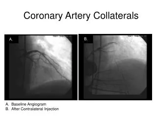
Hypothesis & Study Design • 3 Categories of Infarct Core volume involved in IAT patient selection: • DWI < 70 ml: “Likely to Benefit” • DWI 70-100 ml: “Uncertain to Benefit” • DWI > 100 ml: “Unlikely to Benefit” • Study Design for ”Transfer to Hub” Decision: • “Malignant” vs “Non-malignant” • DEFINITION of “malignant” on CTA = • Absent or markedly decreased collaterals over >50% of MCA territory –OR– • Marked GM/WM hypo-enhancement on CTA with volume > 100 ml • Hypothesis Regarding CTA collaterals status & hypo-enhancement size: • “Likely to Benefit”: “Non-malignant” • “Uncertain to Benefit”: “Non-malignant” • “Unlikely to Benefit”: “Malignant”
Goal & Methods • Comparison between … • 2 different commercially available CTP software platforms (“CTP A” & “CTP B”), vs • 3 different reader levels-of-experience interpretation of CTA collaterals and hypo-enhancement • … for estimation of DWI “core” infarct volume of < or > 100 ml • 3 different reader levels-of-experience: • 2 CAQ-level Staff Neuroradiologists (consensus) • ED Radiology 1st year Fellow • Neuro-IR Fellow (2-yrs neuroimaging experience) • N=55 consecutive patients with anterior circulation acute ischemic stroke who underwent CTA, CTP, & DWI-MRI within 1-hour of presentation. Hypo-enhancing “core” volume on the CTA estimated by: (L x W x H)/2
Methods Patients were stratified by DWI core volume into >100ml and <100ml Sensitivities and specificities were calculated for each CTP software platform and each reader’s CTA score Inter-observer agreement was calculated with the Kappa statistic
Methods: Non-malignant pattern - DWI volume of 74 ml CTA hypo-enhancement volume (L x W x H)/2 = 5.5 x 5 x 5/2 = 69ml DWI volume 74ml
Results Strong inter-observer agreement between the “Consensus CAQ NR” scores with Cohen’s Kappa: 0.89
Conclusion For the purpose of making transfer decisions from “spoke” to “hub” centers for IAT treatment – if DWI is unavailable for accurate “core” assessment - a simple and easy to implement standardized CT/CTA protocol may be sufficient, without added sensitivity or specificity provided by CTP Additional prospective studies with more patients are required for validation of our results