Download
1 / 30
340 likes | 870 Views
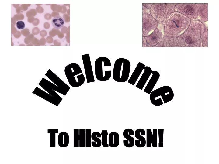
Welcome. To Histo SSN!. Microscopy and Cytology Stains. Intro to Histo Handout (tips on course) Important terms (Acidophilic, Basic, etc.) Examples of some stains you’ll see Vital stain (of blood smear) EMs Any specific questions you have, please hold to the end. Basophilic.

E N D
Welcome To Histo SSN!
Microscopy and Cytology Stains • Intro to Histo Handout (tips on course) • Important terms (Acidophilic, Basic, etc.) • Examples of some stains you’ll see • Vital stain (of blood smear) • EMs Any specific questions you have, please hold to the end.
Basophilic • Basophilic structures are stained by basic dyes: • Basic dyes are positive • Basophilic structures are negative (ex. DNA, RNA, ribosomes, RER) • Mnemonic:Basophilic = Blue Online Reference: Lab #1 Slide #14
Acidophilic (Eosinophilic) • Acidophilic structures are stained by acid dyes: • Acid dyes are negative • Acidophilic structures are positive (ex. Proteins, collagen, cytoplasm) • Eosinophilic = Pink Online Reference: Lab #1 Slide #14
Question 1 • How would you describe the structure at the pointer? • Eosinophilic • Basophilic • Negatively charged • Positively charged Online Reference: Lab #1 Slide #16
Question 1 • How would you describe the structure at the pointer? • Eosinophilic • Basophilic • Negatively charged • Positively charged Online Reference: Lab #1 Slide #16
Nissl Stain Lab #1 Slide #1 Lab #1 Slide #2
Nissl Stain • Blackish blue • Stains negatively charged structures • Nissl substance (bodies) = stacks of RER and free polyribosomes • Nucleolus = site of rRNA synthesis Lab #1 Slide #3
Question 2 • What cellular function is occurring in this region of this cell? • rRNA transcription • RNA translation • Protein degradation • DNA degradation Lab #1 Slide #3
Question 2 • What cellular function is occurring in this region of this cell? • rRNA transcription • RNA translation • Protein degradation • DNA degradation Lab #1 Slide #3
Eosin (H&E) • Pink • Stains Eosinophilic structures ex. Proteins, collagen Lab #1 Slide #13
Hemotoxylin (H&E) • Blue, purple or blackish • Stains Basophilic structures ex. DNA, ribosomes, RNA • Euchromatin is DNA in USE. It is spread out, diffuse, and less stained. • Heterochromatin is condensed DNA, and stains dark blue. Lab #1 Slide #13
Periodic Acid Schiff &Hematoxylin (PAS) • Pink, Magenta • Stains carbohydrates and carb. rich macromolecules ex. Glycogen, mucin, basement membrane, etc. • If you see PAS, think CARBOHYDRATES. Lab #1 Slide #10
H&E vs. PAS Lab #14 Slide #8 Lab #14 Slide #18
Acid Fuschin-Toluidin Blue • Pink and Blue • Pink = acid Fuschin • Blue = Toluidin • Akin to H&E • Note that: • Acid dyes stain apical surface • Basic dyes stain basal surface Lab #1 Slide #5
Question 3 • The pink regions are eosinophilic due to: • Endosomes • rER • Cytosolic proteins • Golgi • Cilia Lab 7 #18
Question 3 • The pink regions are eosinophilic due to: • Endosomes • rER • Cytosolic proteins • Golgi • Cilia Lab 7 #18
Question 4 • The apex of this cell stains positively because of • Ribosomes • Carbohydrates • Fats Lab #14 Slide #18
Question 4 • The apex of this cell stains positively because of • Ribosomes • Carbohydrates • Fats Lab #14 Slide #18
Question 5 • The structure at the pointer • Is composed of heterochromatin • Indicates the cell is NOT transcriptionally active • Is the primary site of ribosomal RNA synthesis • Is a Nissl body Lab 6, #18
Question 5 • The structure at the pointer • Is composed of heterochromatin • Indicates the cell is NOT transcriptionally active • Is the primary site of ribosomal RNA synthesis • Is a Nissl body Lab 6, #18
EM 1Identify structures Nucleus Outer Nuclear Membrane Nucleolus Rough ER
EM1, Q1 Proteins that are made here may end up at which of the following sites: • Outside the cell • Golgi Apparatus • Plasma Membrane • All of the above • None of the above Lab 1, Slide 4
EM1, Q1 Proteins that are made here may end up at which of the following sites: • Outside the cell • Golgi Apparatus • Plasma Membrane • All of the above • None of the above Lab 1, Slide 4
EM2 • Active or inactive cell?
EM2 • Active or inactive cell? • ACTIVE
EM2 Q2 Which structure would appear darker in a Nissl stain? • A • B • Neither. They are both eosinophilic • Neither, they do not stain with Nissl Lab 1 Sl 7 B A
EM2 Q2 Which structure would appear darker in a Nissl stain? • A • B • Neither. They are both eosinophilic • Neither, they do not stain with Nissl Lab 1 Sl 7 B A