Download
1 / 15
150 likes | 314 Views
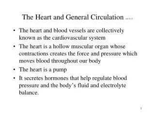
Circulation and The Heart. Chapter 6.2. 6.2.1 Draw and label a diagram of the heart showing the four chambers, associated blood vessels, valves, and the route of blood through the heart. To be done in class on the board.

E N D
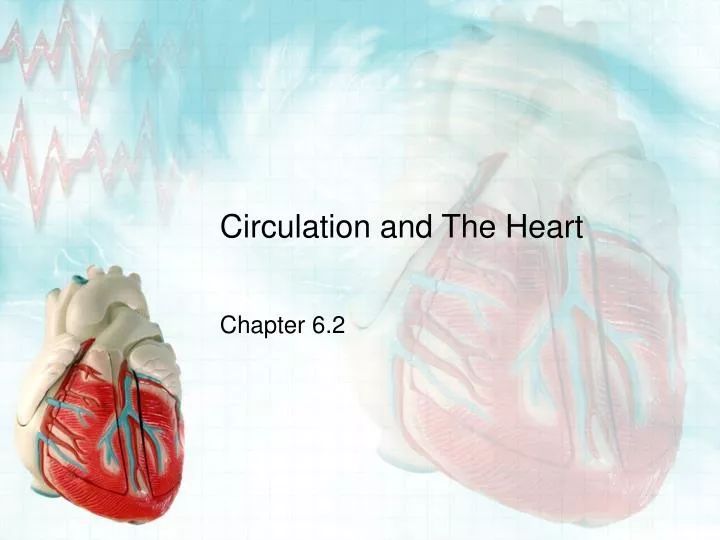
Circulation and The Heart Chapter 6.2
6.2.1 Draw and label a diagram of the heart showing the four chambers, associated blood vessels, valves, and the route of blood through the heart. • To be done in class on the board.
6.2.2 State that the coronary arteries supply heart muscle with oxygen and nutrients. • Coronary arteries branch out into the heart muscle itself and supply the heart with oxygen and nutrients.
6.2.3 Explain the action of the heart in terms of collecting blood, pumping blood, and opening and closing valves. • Oxygenated blood from the lungs is pumped into the left atrium. • Blood is pumped through the atrioventricular valve to push blood into the left ventricle and the AV valve closes. • The left ventricle forces open the semilunar valve to push the blood into the aorta and out into the rest of the body.
6.2.3 Explain the action of the heart in terms of collecting blood, pumping blood, and opening and closing valves. (cont) • Upon its return from the body via the vena cava, the blood enters the right atrium. • The right atrium pumps the blood through the AV valve into the right ventricle. • The AV valve closes and the right ventricle contracts, pumping the blood through a semilunar valve into the pulmonary arteries toward the lungs.
6.2.4 Outline the control of the heartbeat in terms of myogenic muscle contraction, the role of the pacemaker, nerves, the medulla of the brain and adrenaline. • Myogenic muscle contraction: cardiac cells are self-excitable • cardiac muscles can contract without any signal from the nervous system • even though cells of cardiac muscle have an intrinsic ability to contract, they must be coordinated with one another • Pacemaker: controls the rhythm of the heart
6.2.4 Outline the control of the heartbeat in terms of myogenic muscle contraction, the role of the pacemaker, nerves, the medulla of the brain and adrenaline. (continued) • Pacemaker or sinoatrial (SA) node: • in the wall of the right atrium, at superior vena cava junction • made of specialized muscle tissue that combines characteristics of both muscle and nerve • each time the SA node contracts, it starts a quick, wave-like pulse through the walls of the atria • makes the two atria contract in unison http://www.nhlbi.nih.gov/health/dci/Diseases/hhw/hhw_electrical.html
6.2.4 Outline the control of the heartbeat in terms of myogenic muscle contraction, the role of the pacemaker, nerves, the medulla of the brain and adrenaline. (continued) • Atrioventricular (AV) node: • message point for the right atrium and right ventricle • wave of impulse reaches the SV node then has a 0.1 second pause • makes sure that the atria will contract first and be empty before the ventricles contract • after pause, signal travels down to tips of ventricles, then moves up through walls
6.2.4 Outline the control of the heartbeat in terms of myogenic muscle contraction, the role of the pacemaker, nerves, the medulla of the brain and adrenaline. (continued) • Pacemaker is controlled by: • Two sets of nerves that oppose each other in adjusting heart rate – one to speed it up and one top slow it down • Adrenalin: secreted by adrenal gland for ‘fight or flight’ to quicken heart rate • causes the SA node to ‘fire’ more frequently
6.2.4 Outline the control of the heartbeat in terms of myogenic muscle contraction, the role of the pacemaker, nerves, the medulla of the brain and adrenaline. (continued) • Pacemaker is controlled by: • Body temperature: a temperature increase of 1C increases the heart rate by about 10-20 bpm • Exercise: • increases heart rate, due to muscles’ increased need for oxygen • as carbon dioxide levels rise or fall, the medulla senses the CO2 and sends a signal through cranial nerves to the SA node to increase or decrease the heart rate
6.2.5 Explain the relationship between the structure and function of arteries, capillaries and veins. (see table on p.161) 3 kinds of blood vessels • Arteries: carry blood away from the heart • Split into arterioles (smallest type of artery) • Thicker walls for strength because blood flows rapidly at high pressure through the arteries by the heart • Capillaries: super-small vessels with very thin walls to allow exchange • Capillary beds: inside various tissues • Veins: returns blood to the heart • Develop from venules (smallest type of vein) • Thinner walls to deliver blood back to heart at low velocity and pressure
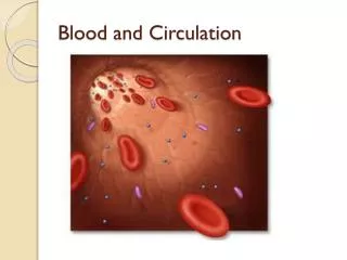
6.2.6 State that blood is composed of plasma, erythrocytes, leucocytes (phagocytes and lymphocytes), and platelets. • Plasma • 55% of blood • Liquid matrix where the components of blood are suspended • Consists of • 90% water • Electrolytes • Proteins (antibodies, clotting factors) • Erythrocytes (red blood cells) • Contain hemoglobin (iron containing protein • Carry O2 and CO2
6.2.6 State that blood is composed of plasma, erythrocytes, leucocytes (phagocytes and lymphocytes), and platelets. • Leucocytes (white blood cells) • Phagocytes • Lymphocytes • Platelets • Cell fragments that assist in blood clotting defense and immunity
6.2.7 State that the following are transported by the blood: nutrients, oxygen, carbon dioxide, hormones, antibodies, urea, and heat. • Function of blood: transport substances throughout the body • Helps maintain stable environment • Contains blood cells and plasma • Transports: • Heat: skin arterioles change diameter – why? • Nutrients: glucose, amino acids • Oxygen: needed for aerobic cellular respiration • Carbon dioxide: waste product of CR • Hormones: transported from gland to target cells • Antibodies: immunity • Waste products: urea