Download
1 / 31
340 likes | 658 Views
The Heart and Circulation. Cardiovascular System = Heart, Blood and Vessels Lymphatic System = Lymph nodes, Organs and Vessels. The Heart. External Innervation Vagus (parasympathetic) C + T sympathetic chain ganglion Pericardium (3 layers) 1) Outer-fibrous pericardium Serous pericardium

E N D
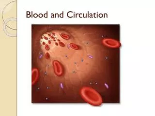
The Heart and Circulation Cardiovascular System = Heart, Blood and Vessels Lymphatic System = Lymph nodes, Organs and Vessels
The Heart • External Innervation • Vagus (parasympathetic) • C + T sympathetic chain ganglion • Pericardium (3 layers) • 1) Outer-fibrous pericardium • Serous pericardium • 2) parietal • 3) visceral (epicardium) • Pericardial Cavity • between layers of serous pericardium • serous fluid • lubricate heart while beating pg 502
Location of Heart in Chest • Oblique Position • Apex = Left of Midline (5th ICS), Anterior to rest of heart • Base (posterior surface) sits on vertebral column • Superior Right = 3rd Costal Cartilage, 1” right midsternum • Superior Left = 2nd Costal Cartilage, 1” left midsternum • Inferior Right = 6th Costal Cartilage, 1” right midsternum • Inferior Left = 5th Intercostal Space at Midclavicular line
Location of Heart in Thorax pg 501
External Features of Heart • Interventricular sulcus • Coronal/Coronary sulcus • Auricles of atria • Apex • Base • Coronary vessels • Ligamentum Arteriosum pg 504
The Great Vessels and major branches Aorta (from Left Ventricle) • Ascending • Coronary arteries • Aortic Arch • Brachiocephalic trunk • Left Common Carotid • Left Subclavian • Descending (Thoracic/Abdominal) • Many small branches to organs Pulmonary Trunk (from Rt Ventricle) • -2 Pulmonary Arteries into lungs Inferior/Superior Vena Cava - Coronary sinus Pg 504, 532
Layers of Heart • Epicardium (most superficial) – Visceral pleura • Myocardium (middle layer) • Cardiac muscle • Contracts • Endocardium (inner) • Endothelium on CT • Lines the heart • Creates the valves pg 502
Right Heart Chambers: Pulmonary Pump • Right Atrium (forms most of base of heart) • Receives O2-poor blood from body via IVC, SVC, Coronary sinus • Ventral wall = rough Pectinate muscle • Fossa Ovalis- on interatrial septum, remnant of Foramen Ovale • Right Ventricle • Receives O2-poor blood from right atrium through tricuspid valve • Pumps blood to lungs via Pulmonary Semilunar Valve in pulmonary trunk • Trabeculae Carnaemuscle ridgesalong ventral surface • Papillary Muscle-cone-shaped muscle to which chordae tendinae are anchored • Moderator Band-muscular band connecting anterior papillary muscle to interventricular septum
Left Heart Chambers: Systemic Pump • Left Atrium • Receives O2-rich blood from 4 Pulmonary Veins • Pectinate Musclesline only auricle • Left Ventricle (forms apex of heart) • Receives blood from Left Atrium via bicuspid valve • Pumps blood into aorta via Aortic Semilunar Valve to body • Same structures as Rt Ventricle: Trabeculae carnae, Papillary muscles, Chordae tendinae • No Moderator Band
Heart Valves: Lub*-Dub** • *Tricuspid Valve: Right AV valve • 3 Cusps (flaps) made of endocardium and CT • Cusps anchored in Rt. Ventricle by Chordae Tendinae • Chordae Tendinae prevent inversion of cusps into atrium • Flow of blood pushes cusps open • When ventricle is in diastole (relaxed), cusps hang limp in ventricle • Ventricular contraction increases pressure and forces cusps closed • *Bicuspid (Mitral) Valve: Left AV valve • 2 cusps anchored in Left Ventricle by chordae tendinae • Functions same as Rt. AV valve • **Semilunar valves: prevents backflow in large arteries • Pulmonary Semilunar Valve: Right Ventricle and Pulmonary Trunk • Aortic Semilunar Valve: Left Ventricle and Aorta • 3 cusps: blood rushes past they’re flattened, as it settles they’re pushed down (valve closed)
Heart Valves pg 506
Flow of Blood • O2-poor blood (S+I VC, Coronary Sinus) enters Rt Atrium • Travels through Tricuspid Valve into Rt Ventricle • Pumped out through Pulmonary Semilunar Valve into Pulmonary trunk (branches into Pulmonary Arteries) and to lungs • After circulating through lungs, O2-rich blood returns to the heart through 4 Pulmonary veins • The O2-rich blood enters the Left Atrium • Travels through Bicuspid/Mitral Valve into Left Ventricle • Pumped out through Aortic Semilunar Valve into Aorta to be distributed to rest of body by descending aorta and branches of aortic arch
Cardiovascular Circuits pg 500
Circuits • Pulmonary Circuit • Vessels carrying blood to and from lungs • Pulmonary arteries and veins • Systemic Circuit • Vessels carrying blood to and from the rest of the body • All other vessels
Blood Flow to Supply the Heart Muscle • Heart wall too thick for diffusion of nutrients • Rt and Lft Coronary Arteries • Branch from Ascending Aorta • Have multiple branches along heart • Sit in Coronary Sulcus • Coronary Heart Disease • Cardiac Veins • Coronary Sinus (largest) • Many branches feed into sinus • Sits in Coronary Sulcus
Anatomy of Arteries and Veins • Tunica externa • Outermost layer • CT w/elastin and collagen • Protects, Strengthens, Anchors • Tunica media • Middle layer • Circular Smooth Muscle • Collagen & Elastic Fibers • Vaso-constriction/dilation • Tunica intima • Innermost layer • Endothelium • Minimize friction • Lumen pg 525
Vessels of Cardiovascular System:Arteries • Carry blood AWAY from heart • Systemic Circuit: carry O2 blood • Pulmonary Circuit: carry de-O2 blood • Walls thicker than Veins • Tunica media > Tunica externa • 3 Types • Conducting (elastic) • large, elastin, high pressure • Distributing (muscular) • medium size, to organs • Arterioles • smallest pg 532
Vessels of Cardiovascular System:Capillaries • Smallest blood vessels • Single layer of endothelium surrounded by basal lamina • Deliver O2 and nutrients to cells and remove waste • Capillary Beds: networks of caps. Regulating amount of blood going to cells throughout tissues • Tendons, Ligaments poorly vascularized • Epithelium, cartilage has no capillaries
Vessels of Cardiovascular System:Veins • Carry blood from capillaries INTO the heart • Systemic Circuit: O2 poor blood • Pulmonary Circuit: O2 –rich blood • Thinner walls than arteries • Tunica externa > tunica media, Less elastin • Larger lumen than arteries • Contain valves (made of T. intima) • Normal movement, Muscular contraction push blood through • Venules- smallest veins
Movement through Veins Pg 529
Cardiovascular Blood Flow • HeartArteries(conducting-distributing) ArteriolesCapillaries of tissues • At Capillaries O2 is delivered and CO2 picked up • CapillariesVenulesVeinsHeart • Portal System: Special vascular circulation where blood goes through 2 capillary beds before returning to the heart to achieve 2nd function • (eg) Hepatic Portal System: aids digestion by picking up digestive nutrients from stomach + intestines and delivers to liver for processing/storage • Pick-up occurs at capillaries of stomach and intestine • Via Hepatic Portal Vein goes to capillaries of liver • Via Hepatic Vein blood goes back to heart
pg 548 Hepatic Portal System
Vascular Anastomoses • Vessels unite and connect • Arteriole Anastomoses • Communication between arteries • Joints, Abdominal Organs, Brain, Heart • Venous Anastomoses • Communication between veins • More common • (eg) back of hand • Vaso Vasorum • Tiny arteries, veins, capillaries in tunica externa of vessels to nourish them (outer half)
1) Fetus must transport blood to and from the placenta 2) Lungs are not functional, and do not need much blood Fetal Circulation: 2 main differences • All major vessels are in place by 3rd month • Blood flows in same direction as in adults
Fetal Circulation: Blood to Placenta • Fetus must supplyplacentaw/blood • Umbilical Vessels:carries blood to/from placenta • 2 Umbilical Arteries= bring blood that contains waste & little O2 from fetus to placenta • 1 Umbilical Vein = brings blood w/O2 and nutrients to fetus from placenta (some goes to portal vein to process in liver) • Ductus Venosus= shunt taking blood returning from placenta to fetus directly to heart, largely bypassing liver • Too much blood for liver to handle • Results in highly O2 blood going to heart
Fetal Circulation: Bypassing the Lungs • Fetal Lungs are not functional and do not need large amounts of blood • Foramen Ovale(becomesFossa Ovalis) • Small hole in inter-atrial septum allows blood to flow directly from Rt. Atrium to Lft. Atrium • This largely bypasses the Rt. VentriclePulmonary trunk that would bring blood to lungs • Ductus Arteriosus (becomesLigamentum Arteriosum) • Shunt directs blood from Pulmonary Trunk to Aortic arch, largely bypassing lungs
Bypassing the Lungs Pg 555
Remnants of Fetal Circulation • Ligamentum teres= Round ligament • Remnant of the umbilical vein • Anterior abdominal wall • Ligamentum venosum • Remnant of ductus venosum • On liver’s inferior surface • Medial Umbilical Ligaments • Remnant of umbilical arteries • Anterior abdominal wall below navel • Also gives branch to urinary bladder
The Lymphatic System • Function: to collect excess tissue fluid collecting at arteriole end of capillary beds, and return leaked blood proteins to blood (maintain osmotic pressure needed to take up water into bloodstream) • Lymph is moved through vessels • Pulse of nearby arteries • Contraction of surrounding skeletal muscle • Regular movement of body (wiggling legs) • Muscle in Tunica Media • Lacteals-lymphatic capillaries w/unique function • In mucosa of small intestine, receive digested fat from intestine • Fatty lymph becomes milky = Chyle • Chyle goes to bloodstream
Lymphatic System…The Players: • Lymph- clear fluid from loose CT at capillaries • Lymphatic capillaries (near blood capillaries) • Lymph collecting vessels (small, 3 tunicas, # valves) • Lymph nodes (sit along collecting vessels)-clean lymph of pathogens, they are NOT glands • Lymphatic trunks (convergence large collecting vessels) • Lumbar, intestinal, bronchomediastinal, subclavian, jugular • Lymphatic ducts empty into veins of neck