Download
1 / 21
390 likes | 823 Views
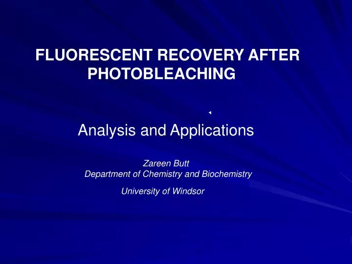
FLUORESCENT RECOVERY AFTER PHOTOBLEACHING Analysis and Applications Zareen Butt Department of Chemistry and Biochemistry University of Windsor. OVERVIEW

E N D
FLUORESCENT RECOVERY AFTER PHOTOBLEACHING Analysis and Applications Zareen Butt Department of Chemistry and Biochemistry University of Windsor
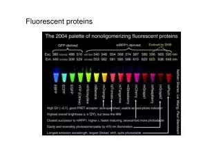
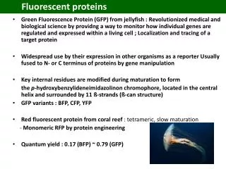
OVERVIEW • Phenomenon of Photobleaching • Fluorescent Recovery After Photobleaching • Measure the mobility of nuclear proteins, macromolecular diffusion within cell membranes, the cytoplasm, nucleoplasm
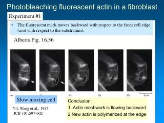
HOW FRAP WORKS • Molecules covalently bound to a fluorophore • Inhomogenous fluorescent population • Spatial separation between fluorescent moleclues and photobleached molecules at time 0
Photobleached Population Example of photobleaching in an indian muntjac fibroblast nucleus expressing ASF/SF2 GFP
DATA COLLECTION 0s 10s 20s 30s 90s
FLUORESCENT RECOVERY CURVE Intensity Intensity Time (seconds)
PREFRAP ANALYSIS • Steady-State distribution in living cells • Artifactual Distributions • Diffused distribution • Formation of large spherical aggregates • Examples of these distributions illustrated for histone deactylase-GFP fusion proteins
Abnormal distribution of GFP-tagged histone deacetylases in mouse 10t1/2 cells transfected with HDAC4-GFP (left) and HDAC3-GFP (right).
DATA NORMALIZATION • The raw data must be normalized in order to compensate for: 1) the background signal in the image 2) the loss of total cellular fluorescence due to photobleaching a subregion of the cell 3) any loss of fluorescence that occurs during the course of collection of recovery time series
DATA ANALYSIS • Diffusion coefficient (measures the rate of movement and represents the mean squared displacement of proteins over time) • Effective diffusion coefficient (does not take into consideration any interaction the proteins might undergo in the process of diffusion)
RECENT ENZYMATIC STUDIES USING FRAP ANALYSIS • gp130/Jak 1 interaction • Kinetics of association and the state of activation of GTPases in phagosomes • Mobility of Glucocorticoid Receptor in the nucleus • Phospholipase C-β2 activity and mode of memebrane interactions in living cells
FRAP ANALYSIS OF gp-130YFP AT THE PLASMA MEMBRANE • Cos-7 cells transfected with a gp130-YFP containing expression vector • Region of interest with a diameter of 1.3µm is photobleached • As a result of double bleaching, the mobile and immobile fractions remains constant
The fraction of mobile and immobile fractions remains constant after double bleaching
FRAP recovery curves demonstarting that Rac 2(12V) reduces fluorescent recovery rate of GFP-PLCß2
RECENT ENZYMATIC STUDIES USING FRAP ANALYSIS • gp130/Jak 1 interaction • Kinetics of association and the state of activation of GTPases in phagosomes • Mobility of Glucocorticoid Receptor in the nucleus • Phospholipase C-β2 activity and mode of memebrane interactions in living cells
CONCLUSION • In the future, FRAP combined with useful mathematical analysis, and use of engineered proteins will serve as an important tool to study the mobility of molecules in living cells
REFERENCES • Carrero, G., Macdonald, D., Crawford, E., Vries de., and Hendzel, M. (2003) Methods. 29, 14-28 • Giese, B., Au-Yeung, C., Herrmann, A., Diefenbach, S., Haan, C., Kuster,A., Wortmann S., Roderburg, C., Heinrich P., Behrmann, I., and Muller-Newen, G. (2003) The journal of biochemistry.278, 39205-39213 • Illenberger, C., Walliser, C., Strobel, J., Gutman, O., Niv, H., Gaidzik, V., Kloog Y., Gierschik, P., and Henis, Y. (2003) The journal of biochemistry. 278, 8645-8652
Schaaf, M., and Cidlowski, J. (2003) Molecular and Cellular Biology. 23, 1922-1934 • Vieira, O., Bucci, C., Harrison, R., Trimble, W., Lanzetti, L., Greunberg J., Schreiber, A., Stahl, P., and Grinstein, S. (2003) Molecular and Cellular Biology.23, 2501-2514