Download
1 / 18
210 likes | 553 Views
X-Rays. Wavelength the size of an atom Frequencies above UV. Uses of X-rays. Used in airports to examine luggage for the presence of dangerous weapons or bombs or for illegal transit of goods.

E N D
X-Rays Wavelength the size of an atom Frequencies above UV
Uses of X-rays • Used in airports to examine luggage for the presence of dangerous weapons or bombs or for illegal transit of goods. • used to detect structural deficits or cracks in metal objects such as bridges and aircraft that are likely to be missed by the human eye • widely used in medicine to reveal the architecture of the bone and other soft tissues and to find out any abnormality in the form of fracture, growth of tumor • also used in dental imaging • Used to determine structure of crystalline chemicals • Used in the study of space
How X-Ray Images are Formed • The atoms that make up your body tissue absorb visible light photons very well. The energy level of the photon fits with various energy differences between electron positions. Radio waves don't have enough energy to move electrons between orbitals in larger atoms, so they pass through most stuff. X-ray photons also pass through most things, but for the opposite reason: They have too much energy. • They can, however, knock an electron away from an atom altogether. Some of the energy from the X-ray photon works to separate the electron from the atom, and the rest sends the electron flying through space. A larger atom is more likely to absorb an X-ray photon in this way, because larger atoms have greater energy differences between orbitals -- the energy level more closely matches the energy of the photon. Smaller atoms, where the electron orbitals are separated by relatively low jumps in energy, are less likely to absorb X-ray photons. • The soft tissue in your body is composed of smaller atoms, and so does not absorb X-ray photons particularly well. The calcium atoms that make up your bones are much larger, so they are better at absorbing X-ray photons. • To see what happens inside an x-ray machine go to http://health.howstuffworks.com/x-ray2.htm
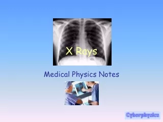
Medical X-Rays • To take an X-ray of the bones, short X-ray pulses are shot through a body with radiographic film behind. The bones absorb the most photons. The X-rays that do not get absorbed turn the photographic film from white to black, leaving a white shadow of bones on the film. • X-rays are especially useful in the detection of pathology of the skeletal system, but are also useful for detecting some disease processes in soft tissue. Some notable examples are the very common chest X-ray, which can be used to identify lung diseases such as pneumonia, lung cancer or pulmonary edema
Medical X-Rays Lung Cancer Broken humerus
Use in Dentistry Dental X-rays spot tooth and tissue damage including abscesses, infections, gum disease and decay
X-Rays in Security Are used to inspect bags before they are placed on airplanes Can also be used to inspect cargo coming in to country through ports
X-Ray Crystallography • The pattern produced by the diffraction of X-rays through the closely spaced lattice of atoms in a crystal is recorded and then analyzed to reveal the nature of that lattice. A related technique, fiber diffraction, was used by Rosalind Franklin to discover the double helical structure of DNA
X-Rays and Astronomy • The Chandra X-ray Observatory, launched on July 23, 1999, has been allowing the exploration of the very violent processes in the universe which produce X-rays. Unlike visible light, which is a relatively stable view of the universe, the X-ray universe is unstable, it features stars being torn apart by black holes, galactic collisions, and novas, neutron stars that build up layers of plasma that then explode into space.
A View of Space • a supernova remnant in the Small Magellanic Cloud. The Chandra X-ray image (blue) shows gas that has been heated to millions of degrees Celsius by a shock wave moving into matter ejected by the supernova. This gas is rich in oxygen and neon. The radio image (red) made with the Australia Telescope Compact Array, traces the outward motion of a shock wave due to the motion of extremely high energy electrons. The optical image (green) made with the Hubble Space Telescope, shows dense clumps of oxygen gas that have 'cooled' to about 30,000 degree Celsius.
Dangers of X-Rays & Gamma Rays • Gamma rays are extremely penetrating. In fact, several inches of lead or even a few feet of concrete are required to stop gamma rays. They are a radiation hazard for the entire body, meaning that although they will pass through you, your tissue will absorb some rays. Gamma rays occur naturally in minerals like potassium-40. • X-rays are essentially the same as gamma rays, but their origin is different. Where gamma rays come from inside the nucleus of an atom, X-rays come from processes outside the nucleus. X-rays come from a change in the electron structure of an atom and are mostly machine-produced. They aren't quite as penetrating as gamma rays, and just a few millimeters of lead can stop them. That's why you wear a "lead apron" when receiving medical X-rays. • Overexposure to ionizing radiation can cause mutations in your genes, which causes birth defects, a raised risk of cancer, burns or radiation sickness
Radiation Doses • In the United States, people receive an average annual dose of about 360 mrem. More than 80 percent of this dose comes from natural background radiation. • Where and how you live affects the amount of radiation exposure you receive. For example, people who live in the Pacific Northwest part of the United States typically only receive about 240 mrem from natural and man-made sources. However, people in the Northeast receive up to 1700 mrem per year, mostly due to radon that is natural to rocks and soil.
Typical Doses of Radiation • We absorb radiation from a variety of sources. How much is too much? Experts say 3 mSv per year is probably OK for most of us; 20 mSv for those who must have medical tests. • RADIATION AMOUNT* • CT scan, full body 10–12 mSv • CT scan, chest or pelvis 4–8 mSv • Natural background radiation (from sunlight, radon gas, etc.) from living in high-altitude cities (e.g., Denver, Salt Lake City) 6 mSv (per year) • Natural background radiation from living at sea level (e.g., Chicago) 3 mSv (per year) • Mammogram 1–2 mSv • High-mileage frequent flying (100,000–450,000 miles per year) 1–6.7 mSv • X-ray of chest (or ankle to look for broken bones) 0.1–0.6 mSv • DEXA (bone-density) scan 0.01–0.05 mSv • Dental X-ray (bitewing) 0.02 mSv • Single airplane flight, coast-to-coast 0.01–0.03 mSv • *mSv=millisievert, the scientific unit of measurement for radiation dose. At high levels, radiation can mutate the structure (genetic components) of a body’s dividing or reproducing cells and increase cancer risks. Sources: American College of Radiology; Radiological Society of North America; American Association of Physical Medicine; The New England Journal of Medicine; University of California, San Francisco, Cancer Center.
Gamma Rays • Very short wavelength (size of atomic nuclei) • Extremely high frequencies and energy • Can penetrate up to 3 m of concrete • Generated by radioactive atoms and in nuclear explosions
Uses of Gamma Rays • Used to study space • Used in medicine for imaging and cancer treatment • To kill germs (irradiated food) • To sterilize medical equipment • To look inside large cargos (security) • Reveal information about the structure of the atomic nucleus
Gamma Rays in Medicine • Because Gamma rays can kill living cells, they are used to kill cancer cells without having to resort to difficult surgery. • This is called "Radiotherapy", and works because cancer cells can't repair themselves like healthy cells can when damaged by gamma rays. Getting the dose right is very important! • There's also targeted radiotherapy, where a radioactive substance is used to kill cancer cells - but it's a substance that'll be taken up by a specific part of the body, so the rest of the body only gets a low dose. An example would be using radioactive iodine to treat cancer in the thyroid gland.
One Tool- The Gamma Knife • The Gamma Knife is faster and more precise than other radiosurgical tools that are currently available. It uses 201 separate radiation beams to target lesions that have been defined by MRI scans or angiograms. The 201 individual beams intersect at a single spot with the accuracy of less than one-tenth of a millimeter (about the thickness of a sheet of paper). Referred to as "surgery without a scalpel," the Gamma Knife procedure does not require the surgeon to make an incision in the scalp, nor an opening in the skull.
Acoustic neuroma Acoustic neuroma • Before Gamma knife after Gamma knife