Download
1 / 20

E N D
X-Rays The Professional Development Service for Teachers is funded by the Department of Education and Science under the National Development Plan
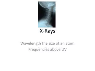
The Electromagnetic Spectrum You know more about this than you may think! The Electromagnetic spectrum or EMS is the name given to all of the types of “waves” which exist in the universe. They all have some things in common e.g. They all travel at 300,000,000 metres per second (3 x 10⁸ ms-1) also they do not have any mass, i.e. they have no weight or atomic structure. The types of waves that people are most familiar with are visible light (light we can see), infra red light, ultra violet light, x rays, micro-waves and radio waves
Gamma Rays are a type of ray emitted by radioactive materials. They are extremely hazardous to living things, but when used correctly they can kill cancer cells. Wavelength ≈ 1x10-12m Frequency 1x10²¹Hz This is the light Humans can see, it makes up the colours of the rainbow, Red, Orange, Yellow, Green, Blue, Indigo, Violet. Wavelength ≈ 1x 10⁻⁶ m Frequency ≈1 x 10¹⁴ Hertz Microwaves, commonly used in cooking and mobile phones, Frequency ≈ 1 x 10⁹ Hz Wavelength ≈1m X- Rays : Used a lot in medicine to help doctors see broken bones, x-rays can pass through tissue but not bone. Wavelength ≈ 1 x 10⁻⁹m Frequency ≈ 1 x 10¹⁸ Hz Infra red, we cant see it , but we feel its effects…its called HEAT. Wavelength≈ 1x 10⁵m, Frequency ≈ 1 x 10¹¹Hz Ultra Violet.. This is responsible for people getting a tan or sunburn!! Wavelength ≈ 1 x 10⁻⁷m Frequency ≈ 1 x 10¹⁶Hz
X-Rays • This unit deals with X-rays. By the end of this unit you should be able to explore and explain some of the following ideas • Some applications of x-rays in medicine. • Some background information about x-rays. • Some of the physics ideas behind x-rays. • Complete a short experiment to show a simulation of how x-rays work. • Write an explanation of how your experiment shows how x-rays work. • 6. Have the basic knowledge needed for Expert Group tasks
X-RaysMedical Applications X-rays are used in medicine for medical analysis. Dentists use them to find complications, cavities and impacted teeth. Soft body tissue are transparent to the waves. Bones and teeth block the rays and show up as white on the black background
Below are some x-rays showing objects which have been swallowed by people. The examples show an open safety pin and a child's stomach with all the pieces of a magnetic toy re-aligned after he's swallowed them…one by one!
X-Rays and Mammograms A mammogram (also called a mammography exam) is a safe, low-dose x-ray of the breast. A high-quality mammogram is the most effective tool for detecting breast cancer early. Early detection of breast cancer may allow more treatment options. It could even mean saving a woman’s breast or her life.
How is a mammogram taken? Using a low-dose x-ray, the mammogram machine takes a snapshot of the inside of a woman’s breast. The machine holds and compresses the breasts so that images at different angles can be taken. Doctors and nurses examine these snapshots, looking for signs of abnormalities such as lumps, which could be tumors. The results of a mammogram are usually available rather quickly, easing the anxiety of those undergoing the procedure
A mammogram showing a healthy breast (left), and a mammogram showing cancer (right). Breast x-rays can detect breast lumps or changes long before they can be felt. The information provided by a mammogram can lead to proper follow-up care. Healthy Cancerous
History of X-Rays As with many of mankind's monumental discoveries, x-ray technology was invented completely by accident. In 1895, a German physicist named Wilhelm Roentgen made the discovery while experimenting with electron beams in a gas discharge tube. Roentgen noticed that a fluorescent screen in his lab started to glow when the electron beam was turned on. Wilhem Roentgen, The discoverer of X-Rays
Roentgen placed various objects between the tube and the screen, and the screen still glowed. Finally, he put his hand in front of the tube, and saw the silhouette of his bones projected onto the fluorescent screen. So he not only discovered x-rays but also, their most beneficial application. One of the very first x-rays: The picture above shows an x-ray taken of Wilhem Roentgen wife's hand. You can see her wedding ring is clearly visible.
Roentgen's remarkable discovery was one of the most important medical advancements in human history. X-ray technology lets doctors see straight through human tissue to examine broken bones, cavities and swallowed objects with extraordinary ease. Modified x-ray procedures can be used to examine softer tissue, such as the lungs, blood vessels or the intestines. You will find out more on about this when you study radioisotopes. Above: X-Ray of Blood Vessel Below: X-Ray of Lungs
First of all, how do x-rays work? Well, they don't, of course, any more than light 'works'. X-rays describe radiation which is part of the electromagnetic spectrum (EMS). X – Rays have a very short wavelength, generally the shorter the wavelength, the more penetrating the wave. The point about x-rays is that, unlike visible light, they pass straight through some materials, including the materials that people are made of. So, when a person is exposed to an x-ray, and a piece of film is placed on the other side of them, a shadow of the inside of their body is produced. This only works at all because the different tissues that make up our bodies absorb x-rays to different extents. The problem is, that we consist largely of water, so most of our soft bits look much the same as far as x-rays are concerned. In fact, there are really only three tissues that are sufficiently different from each other in terms of x-ray absorption to show up on an x-ray film: bone, air and soft tissue. These show up as grey/white areas on an x-ray. The Physics of X - Rays
How are X-Rays Produced? X-Rays are produced in a special type of tube called… “An X ray Tube!!”
Production Of X-Rays.. (continued) Electrons are first emitted from a heated filament, by a process called thermionic emission. They are then accelerated across the evacuated X-ray tube, under the action of a large voltage across the tube, the filament forming the negative cathode and the target being positive anode. On striking the target, the electrons lose most (about 99%) of their energy in low-energy collisions with target atoms, resulting in a substantial heating of the target. The rest of the electron energy (usually less than 1%) reappears as X-ray radiation.
Production Of X-Rays.. (continued) • A rapidly-rotating anode is generally used. It forms the (tungsten) target surface on to which the electron beam is focused. The target area under bombardment is constantly changing, thus reducing local heat concentration. (You can often hear the whirring of the anode motor during the taking of an X-ray. • Copper, being an excellent heat conductor, is used to hold the anode in place. • Oil, which circulates in the outer housing,, helps with convective cooling (as well as providing electrical insulation).
Experiment to show how X-Rays Work Because x-rays can damage human tissue severely, it is not possible to do a hands-on activity with this type of radiation. There is a "simulated" x-ray activity included in the next slide.
The effect of X rays can be simulated by the following activity: Place a piece of thin wire gauze over a box. Place a pattern made of cardboard on top of the screen. Sprinkle sand over the area of the box. The sand (x-rays) will pass through the screen to the bottom of the box, except where they are blocked by the pattern An outline of the pattern can be seen in the bottom of the box. The photographs show the set up and an example of the pattern in the bottom of the box. An area of no sand shows the shape of the blocking pattern.
Patent? Dr. Roentgen refused to patent any of the apparatus associated with the production of x-rays believing that this new discovery should be used for "the good of man". If the discovery were made today, do you think that the discoverer would be likely to make the same decision?