Download
1 / 39

E N D
Major Histocompatibility Complex (MHC) and T Cell Receptors M.Prasad Naidu MSc Medical Biochemistry, Ph.D.,Research scholar
Historical Background • Genes in the MHC were first identified as being important genes in rejection of transplanted tissues • Genes within the MHC were highly polymorphic • Studies with inbred strains of mice showed that genes within the MHC were also involved in controlling both humoral and cell-mediated immune responses • Responder/Non-responder strains
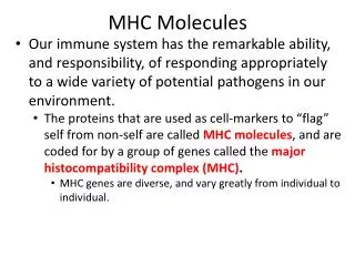
Historical Background • There were three kinds of molecules encoded by the MHC • Class I • Class II • Class III • Class I MHC molecules are found on all nucleated cells (not RBCs) • Class II MHC molecules are found on APC • Dendritic cells, Macrophages, B cells, other cells
Nucleated cells Class I MHC RBCs Class II MHC APCs Historical Background
Historical Background • Class III MHC molecules • Some complement components • Transporter proteins
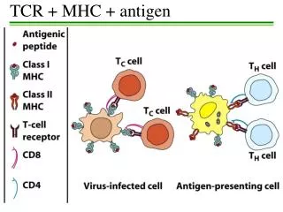
Historical Background • It was not until the discovery of how the TCR recognizes antigen that the role of MHC genes in immune responses was understood • TCR recognizes antigenic peptides in association with MHC molecules • T cells recognize portions of protein antigens that are bound non-covalently to MHC gene products • Tc cells recognize peptides bound to class I MHC molecules • Th cells recognize peptides bound to class II MHC molecules
Historical Background • Three dimensional structures of MHC molecules and the TCR have been determined by X-ray crystallography
Structure of Class I MHC • Two polypeptide chains, a long α chain and a short β (β2 microglobulin) • Four regions • Cytoplasmic region containing sites for phosporylation and binding to cytoskeletal elements • Transmembrane region containing hydrophobic amino acids
Structure of Class I MHC • Four regions • A highly conserved α3 domain to which CD8 binds • A highly polymorphic peptide binding region formed from the α1 and α2 domains • Β2-microglobulin helps stabilize the conformation
Structure of Class I MHC Variability map of Class 1 MHC α Chain
From Janeway et al., Immunobiology 6th Ed. Structure of Class I MHCAg-Binding Groove • Groove composed of an α helix on two opposite walls and eight β-pleated sheets forming the floor • Residues lining the groove are most polymorphic • Groove accomodates peptides of 8-10 amino acids long
From Janeway et al., Immunobiology 6th Ed. Structure of Class I MHCAg Binding Groove • Specific amino acids on peptide required for “anchor site” in groove • Many peptides can bind • Vaccine development
Structure of Class II MHC • Two polypeptide chains,α and β, of roughly equal length • Four regions • Cytoplasmic region containing sites for phosporylation and binding to cytoskeletal elements
Structure of Class II MHC • Four regions • Transmembrane region containing hydrophobic amino acids • A highly conserved α2 and a highly conserved β2 domains to which CD4 binds • A highly polymorphic peptide binding region formed from the α1 and β1 domains
Structure of Class II MHC Variability map of Class2 MHC β Chain
From Janeway et al., Immunobiology 6th Ed. Structure of Class I MHCAg-Binding Groove • Groove composed of an α helix on two opposite walls and eight β-pleated sheets forming the floor • Both the α1 and β1 domains make up the groove • Residues lining the groove are most polymorphic
From Janeway et al., Immunobiology 6th Ed. Structure of Class I MHCAg-Binding Groove • Groove is open and accomodates peptides of 13-25 amino acids long, some of which are ouside of the groove • Anchor site rules apply
Important Aspects of MHC • Although there is a high degree of polymorphism for a species, an individual has maximum of six different class I MHC products and only slightly more class II MHC products (considering only the major loci). • Each MHC molecule has only one binding site. The different peptides a given MHC molecule can bind all bind to the same site, but only one at a time.
Important Aspects of MHC • Because each MHC molecule can bind many different peptides, binding is termed degenerate. • MHC polymorphism is determined only in the germline. There are no recombinational mechanisms for generating diversity. • MHC molecules are membrane-bound; recognition by T cells requires cell-cell contact.
Important Aspects of MHC • Alleles for MHC genes are co-dominant. Each MHC gene product is expressed on the cell surface of an individual nucleated cell. • A peptide must associate with a given MHC of that individual, otherwise no immune response can occur. That is one level of control.
Important Aspects of MHC • Mature T cells must have a T cell receptor that recognizes the peptide associated with MHC. This is the second level of control. • Cytokines (especially interferon-γ) increase level of expression of MHC.
Important Aspects of MHC • Peptides from the cytosol associate with class I MHC and are recognized by Tc cells . Peptides from within vesicles associate with class II MHC and are recognized by Th cells. • Why so much polymorphism? • Survival of the species
Structure of the T cell Receptor • Heterodimer with one α and one β chain of roughly equal length • A short cytoplamic tail not capable of transducing an activation signal • A transmembrane region with hydrophobic amino acids
Structure of the T cell Receptor • Both α and β chains have a variable (V) and constant (C) region • V regions of the α and β chains contain hypervariable regions that determine the specificity for antigen
Structure of the T cell Receptor • Each T cell bears TCRs of only one specificity (allelic exclusion)
Genetic Basis for Receptor Generation • Generation of a vast array of BCRs is accomplished by recombination of various V, D and J gene segments encoded in the germline • Generation of a vast array of TCRs is accomplished by similar mechanisms • TCR β chain genes have V, D and J gene segments • TCR α chain genes have V and J gene segments
Germline ß-Chain Gene Jß11--------Jß16 Cß1 Cß1 Dß2 Dß2 Dß2 Jß11---------------Jß17 Jß11---------------Jß17 Jß11---------------Jß17 Cß2 Cß2 Cß2 Dß1 L L L Vß1 Vß1 Vß1 L L L Vß2 Vß2 L L Vßn Vßn P P P P P P P P D-J rearrangement Dß1Jß15 DNA V-D rearrangement Cß1 Vß2Dß1Jß15 DNA E E E Transcription Cß1 Vß2Dß1Jß15 RNA Organization and Rearrangement of the T Cell Receptor
γδ TCR • Small population of T cells express a TCR that contain γ and δ chains instead of α and β chains • The Gamma/Delta T cells predominate in the mucosal epithelia and have a repertoire biased toward certain bacterial and viral antigens • Genes for the δ chains have V, D and J gene segments; γ chains have V and J gene segments • Repertoire is limited
γδ TCR • Gamma/Delta T cells can recognize antigen in an MHC-independent manner • Gamma/Delta T cells play a role in responses to certain viral and bacerial pathogens
TCR and CD3 Complex • TCR is closely associated with a group of 5 proteins collectively called the CD3 complex • γ chain • δ chain • 2 ε chains • 2 ξ chains • CD3 proteins are invariant
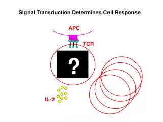
Role of CD3 Complex • CD3 complex necessary for cell surface expression of TCR during T cell development • CD3 complex transduces signals to the interior of the cells following interaction of Ag with the TCR
The “Immunological Synapse” • The interaction between the TCR and MHC molecules are not strong • Accessory molecules stabilize the interaction • CD4/Class II MHC or CD8/Class I MHC • CD2/LFA-3 • LFA-1/ICAM-1
The “Immunological Synapse” • Specificity for antigen resides solely in the TCR • The accessory molecules are invariant • Expression is increased in response to cytokines
The “Immunological Synapse” • Engagement of TCR and Ag/MHC is one signal needed for activation of T cells • Second signal comes from costimulatory molecules • CD28 on T cells interacting with B7-1 (CD80) or B7-2 (CD86) • Others • Costimulatory molecules are invariant • “Immunological synapse”
Costimulation is Necessary for T Cell Activation • Engagement of TCR and Ag/MHC in the absence of costimulation can lead to anergy • Engagement of costimulatory molecules in the absenece of TCR engagement results in no response • Activation only occurs when both TCR and costimulatory molecules are engaged with their respective ligands • Downregulation occurs if CTLA-4 interacts with B7 • CTLA-4 send inhibitory signal
Key Steps in T cell Activation • APC must process and present peptides to T cells • T cells must receive a costimulatory signal • Usually from CD28/B7 • Accessory adhesion molecules help to stabilize binding of T cell and APC • CD4/MHC-class II or CD8/MHC class I • LFA-1/ICAM-1 • CD2/LFA-3 • Signal from cell surface is transmitted to nucleus • Second messengers • Cytokines produced to help drive cell division • IL-2 and others