Download
1 / 83
830 likes | 834 Views
Good Morning!. Please pick up ch.12 notes : cut out and glue into your notebooks, thank you . Agenda. Glue new ch.12 notes into notebooks Lecture/Notes on section 1 Foldable Vocabulary Cards DNA video No homework tonight . 12–1 DNA Griffith and Transformation

E N D
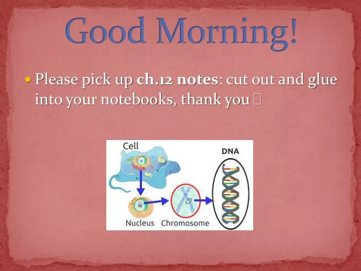
Good Morning! • Please pick up ch.12 notes: cut out and glue into your notebooks, thank you
Agenda • Glue new ch.12 notes into notebooks • Lecture/Notes on section 1 • Foldable • Vocabulary Cards • DNA video • No homework tonight
12–1 DNA Griffith and Transformation • Griffith’s Experiment • Transformation Avery and DNA The Hershey-Chase Experiment • Bacteriophages • Radioactive Markers The Structure of DNA • Chargaff’s Rules • X-Ray Evidence • The Double Helix
DNA • How do genes work? What are they made of, and how do they determine the characteristics of organisms? Are genes single molecules, or are they longer structures made up of many molecules? In the middle of the 1900s, questions like these were on the minds of biologists everywhere. • To truly understand genetics, biologists first had to discover the chemical nature of the gene. If the structures that carry genetic information could be identified, it might be possible to understand how genes control the inherited characteristics of living things.
Griffith’s Experiment • In 1928, British scientist Frederick Griffith was trying to figure out how bacteria make people sick. • Griffith had isolated two slightly different strains, or types, of pneumonia bacteria from mice. The disease-causing strain of bacteria grew into smooth colonies on culture plates, whereas the harmless strain produced colonies with rough edges. The differences in appearance made the two strains easy to distinguish.
When Griffith injected mice with the disease-causing strain of bacteria, the mice developed pneumonia and died. When mice were injected with the harmless strain, they didn’t get sick at all. Griffith wondered if the disease-causing bacteria might produce a poison. • To find out, he took a culture of these cells, heated the bacteria to kill them, and injected the heat-killed bacteria into mice. The mice survived, suggesting that the cause of pneumonia was not a chemical poison released by the disease-causing bacteria.
Transformation • Griffith’s next experiment produced an amazing result. He mixed his heat-killed, disease-causing bacteria with live, harmless ones and injected the mixture into mice. By themselves, neither should have made the mice sick. But to Griffith’s amazement, the mice developed pneumonia and many died. When he examined the lungs of the mice, he found them filled with the disease-causing bacteria. • Somehow the heat-killed bacteria had passed their disease-causing ability to the harmless strain. Griffith called this process transformation because one strain of bacteria (the harmless strain) had apparently been changed into another (the disease-causing strain).
Griffith’s Experiment Heat-killed, disease-causing bacteria (smooth colonies) Harmless bacteria (rough colonies) Harmless bacteria (rough colonies) Control(no growth) Heat-killed, disease-causing bacteria (smooth colonies) Disease-causing bacteria (smooth colonies) Dies of pneumonia Dies of pneumonia Lives Lives Live, disease-causingbacteria (smooth colonies)
Avery & DNA • In 1944, a group of scientists led by Canadian biologist Oswald Avery at the Rockefeller Institute in New York decided to repeat Griffith’s work. They did so to determine which molecule in the heat-killed bacteria was most important for transformation. • Avery and his colleagues made an extract, or juice, from the heat-killed bacteria. They then carefully treated the extract with enzymes that destroyed proteins, lipids, and carbohydrates. Transformation still occurred. Obviously these molecules were not responsible for the transformation. If they had been, transformation would not have occurred, because the molecules would have been destroyed by the enzymes.
Avery and the other scientists repeated the experiment, this time using enzymes that would break down DNA. When they destroyed the nucleic acid DNA in the extract, transformation did not occur. • Avery and other scientists discovered that DNA is the nucleic acid that stores and transmits the genetic information from one generation of an organism to the next.
Hershey-Chase Experiment Bacteriophage with phosphorus-32 in DNA Phage infectsbacterium Radioactivity inside bacterium Bacteriophage with sulfur-35 in protein coat Phage infectsbacterium No radioactivity inside bacterium
Hershey-Chase Experiment Bacteriophage with phosphorus-32 in DNA Phage infectsbacterium Radioactivity inside bacterium Bacteriophage with sulfur-35 in protein coat Phage infectsbacterium No radioactivity inside bacterium
Hershey-Chase Experiment Bacteriophage with phosphorus-32 in DNA Phage infectsbacterium Radioactivity inside bacterium Bacteriophage with sulfur-35 in protein coat Phage infectsbacterium No radioactivity inside bacterium
Hershey-Chase Experiment • 1952: Two American scientists, Alfred Hershey and Martha Chase, studied viruses- nonliving particles smaller than a cell that can infect living organisms. • A virus that infects and kills bacteria is known as a bacteriophage; composed of a DNA or RNA core and a protein coat. • Hershey and Chase reasoned that if they could determine which part of the virus—the protein coat or the DNA core—entered the infected cell, they would learn whether genes were made of protein or DNA.
To do this, they grew viruses in cultures containing radioactive isotopes of phosphorus-32 (32P) and sulfur-35 (35S). The radioactive substances could be used as markers. If 35S was found in the bacteria, it would mean that the viruses’ protein had been injected into the bacteria. If 32P was found in the bacteria, then it was the DNA that had been injected. • All the radioactivity in the bacteria was from phosphorus (32P), the marker found in DNA. Hershey and Chase concluded that the genetic material of the bacteriophage was DNA, not protein.
DNA Scientists Foldable • Fold a piece of your notebook paper toward the spiral. • Cut top part into 3 equal sections. • Label the outside of the 3 sections with the 3 Scientists. • Write the experiment they did and what their conclusion was on the inside paper. 5 minutes
DNA Structure • 3 Functions of DNA: • Genes carry information from one generation to the next. • They had to put that information to work by determining the heritable characteristics of organisms. • Genes had to be easily copied. • DNA is a long molecule made up of subunits called nucleotides. • Each nucleotide is made up of 3 parts: a 5-carbon sugar called deoxyribose, a phosphate group, and a nitrogen base. • There are 4kinds of nitrogenous bases in DNA. Two of the nitrogenous bases, adenine (A) and guanine (G), belong to a group of compounds known as purines. The remaining two bases, cytosine (C) and thymine (T), are known as pyrimidines. • Purines have two rings, and pyrimidines have one ring.
The backbone of a DNA chain is formed by sugar and phosphate groups of each nucleotide. The nitrogenous bases stick out sideways from the chain. The nucleotides can be joined together in any order, meaning that any sequence of bases is possible. • Chargaff’s Rule: Erwin Chargaff, an American biochemist, had discovered that the percentages of guanine [G] and cytosine [C] bases are almost equal in any sample of DNA. The same thing is true for the other two nucleotides, adenine [A] and thymine [T].
X-Ray Evidence • 1950: A British scientist named Rosalind Franklin began to study DNA. She used a technique called X-ray diffraction to get a picture of the structure of a DNA molecule. • By itself, Franklin’s X-ray pattern doesn’t reveal the structure of DNA. The X-shaped pattern in the image does shows that there are 2 strands in DNA that are twisted around each other, a shape known as a helix.
The Double Helix • At the same time that Franklin was continuing her research, Francis Crick, a British physicist, and James Watson, an American biologist, were trying to understand the structure of DNA by building three-dimensional models of the molecule. • 1953: Watson was shown a copy of Franklin’s remarkable X-ray pattern and reported the pattern’s clues to Crick. • Within a few weeks, Watson & Crick figured out the final structure of DNA: a two stranded double helix.
Watson & Crick discovered that hydrogen bonds could form between certain nitrogenous bases and provide just enough force to hold the two strands together. • Hydrogen bonds can form only between certain base pairs—adenine & thymine and guanine & cytosine. Once they saw this, it explained Chargaff’s rules. A = T and G = C. Nucleotide Hydrogen bonds Sugar-phosphate backbone Key Adenine (A) Thymine (T) Cytosine (C) Guanine (G)
Summary • Please spend 2 minutes writing a summary about what we have learned from ch.12 section 1.
Vocabulary Cards In your groups, write the definition in your own words, and add an example and/or a picture. • DNA Structure • 3 Functions of DNA • Griffith • Avery • Hershey-Chase • Chargaff’s Rule • Rosalind Franklin • Watson & Crick • Purine & Pyrimidine 3 minutes
Warm-up • Please pick up 1 warm-up card per pair • Glue in new notes for 12-2 also!
Agenda • Warm-up cards from section 1 • Lecture/Notes on section 2 • Vocabulary Cards • DNA Replication Activity
12–2 Chromosomes & DNA Replication DNA and Chromosomes • DNA Length • Chromosome Structure DNA Replication • Duplicating DNA • How Replication Occurs
DNA & Chromosomes • Prokaryotic cells do not have a nucleus, instead, their DNA is located in the cytoplasm and consists of a single circular chromosome of genetic information.
Eukaryotes have 1,000 times more DNA than prokaryotes! Eukaryotic DNA is found in the nucleus and forms into many chromosomes. • Humans have 46 chromosomes, fruit flies have 8, and trees have 22. • DNA Length: the single chromosome of a prokaryotic E. coli bacteria contains 4 million base pairs (letters), and is about 1.6mm long. DNA has to be tightly folded to fit inside of a tiny cell. • Eukaryotic DNA contains 30 million base pairs, and is 10x longer than a bacteria's chromosome. Eukaryotic chromosomes contain both DNA and proteins (histones) that help it to stick together as tightly packed nucleosome coils.
Chromosome Structure of Eukaryotes Chromosome Nucleosome DNA double helix Coils Supercoils Histones
DNA Replication • DNA stands are complementary, meaning that each side matches the other: chargaff’s rule, so DNA can be easily copied by following the base pairing rule. • Before cells divide (mitosis) their DNA must be duplicated in a process called DNA Replication. • DNA Replication: the DNA molecule separates into 2 stands, then produces 2 new complementary strands following the rules of base pairing. Each strand of the double helix of DNA serves as a template/model for the new stands.
How DNA Replication Occurs • DNA replication is carried out by enzymes. Remember that enzymes help speed up reactions and are very specific (lock & key). • The enzyme Helicase unzips a molecule of DNA first by breaking the hydrogen bonds between the base pairs. Each strand serves as a template for the attachment of complementary bases. • The enzyme DNA Polymerase adds new base pairs to the new DNA stand, and also proofreads to make sure it is adding the correct bases to make a perfect copy.
DNA Replication Original strand New strand DNA polymerase Growth DNA polymerase Growth Replication fork Replication fork Nitrogenous bases New strand Original strand
Each DNA molecule resulting from replication has 1 original strand and 1 new stand. • Example: a strand that is TACGTT produces a complementary stand with bases ATGCAA.
Summary • Please spend 2 minutes writing a summary about what we have learned from ch.12 section 2.
Vocabulary Cards • In your groups, write the definition in your own words, and add an example or a picture. • Prokaryotic Chromosome • Eukaryotic Chromosomes • DNA Replication • Mitosis • Helicase • DNA Polymerase
DNA Replication Activity • In pairs: • Pick up 1 DNA Replication Packet • Read through the first page together, then follow the directions on the second page. • Take turns with different steps. • When finished, each person glues ½ of the replicated copy of DNA into their notebook. • Homework: answer the 6 questions at the end 30 minutes
Warm-up • Please pick up 1 card EACH • Glue in the diagram for today’s notes.
Agenda • Notes on 12-3 • Foldable • Transcription/Translation Practice • 12-3 Summary Videos • Snork DNA Activity • Homework due next class.
12–3 RNA and Protein Synthesis The Structure of RNA • Types of RNA Transcription • RNA Editing • The Genetic Code Translation
RNA & Protein Synthesis • The double helix structure of DNA shows how easily it can be replicated, but it doesn't explain how a gene works to give us genic information. • Genes are coded DNA instructions that control the production of proteins within the cell. The first step in decoding these genetic messages is to copy part of the nucleotide (base pair) sequence from DNA into RNA. • The RNA molecules are then used to make proteins.
Structure of RNA • RNA, like DNA, consists of a long chain of nucleotides. Remember, nucleotides are made of 3 parts: a 5-carbon sugar, a phosphate, and a nitrogen base. • Three differences between RNA and DNA: • RNA contains the sugar “ribose”, where DNA contains the sugar “deoxyribose”. • RNA is a single stand, and DNA is a double strand. • RNA contains the bases A,C, G, U; and DNA contains the bases A, C, G, T. • You can think of RNA as a disposable copy of a segment of DNA that is only used as 1 time instructions for a single gene.
Types of RNA • There are 3 types of RNA that help with protein synthesis: • mRNA (messenger RNA = the single stand of code) • rRNA (ribosomal RNA = what ribosomes are made of) • tRNA(transfer RNA = brings amino acids to the ribosome in the order of the mRNA code) rRNA tRNA mRNA
MessengerRNA Ribosomal RNA Transfer RNA Bringamino acids toribosome Combine with proteins tRNA mRNA Carry instructions rRNA DNA Ribosome Ribosomes RNA can be also called which functions to also called which functions to also called which functions to from to to make up
Foldable • Fold paper in ½ towards the bottom of your notebook. • Cut 2 vertical sections to make 3 areas to write: • Outside: list & draw the 3 types of RNA. • Inside: Describe what their job and location.
Transcription Adenine (DNA and RNA) Cystosine (DNA and RNA) Guanine(DNA and RNA) Thymine (DNA only) Uracil (RNA only) RNApolymerase DNA RNA
Transcription • A process in the nucleus, where mRNA is made by copying part of the nucleotide sequence (a gene) in DNA. Transcription requires an enzyme called RNA Polymerase (acts just like DNA Polymerase by adding RNA nucleotides: A, U, G, C). • How does RNA Polymerase know where to start? There is a region on DNA known as a Promoter, which signals where to bind and start transcription.
RNA Editing • Like a writer’s first draft of a paper, RNA molecules require editing before they are ready to leave the nucleus and make protein. • There are 2 main parts to mRNA: • Introns: “in between” pieces that are not needed. • Exons: “exactly” the genes to be read to make protein. • The introns get cut out and we are left with only exons in our mRNA strand. It can now leave the nucleus and go to a ribosome to be read.
Reading RNA • Proteins are made by joining amino acids together into chains called polypeptides. There are 20 different types of amino acids. • By reading the nucleotide sequence of mRNA, we can “translate” the letters into amino acids, and join them together one after another to make a protein. • We read 3 mRNA letters at a time to make 1 amino acid: each set of 3 letters is called a Codon. • Ex: UCGCACGGU would be read as: UCG-CAC-GGU, and would make the amino acids: serine-histidine-glycine