Download
1 / 59
600 likes | 732 Views
Virus of Fungi, Yeast and Parasitic Microorganism. Anggota kelompok : Waode Nur Zara 104070 30 Yunitazari Laksmi PW 10407031 Venessa Alia 104070 32 Siti Amalia 104070 33 Sutina 104070 34 Vilandri Astarini 10407035 Angga Kusnan Qodafi 104070 36 Oktira Roka Aji 10407038

E N D
Virus of Fungi, Yeast and Parasitic Microorganism.
Anggota kelompok : • Waode Nur Zara 10407030 • Yunitazari Laksmi PW 10407031 • Venessa Alia 10407032 • Siti Amalia 10407033 • Sutina 10407034 • Vilandri Astarini 10407035 • Angga Kusnan Qodafi 10407036 • Oktira Roka Aji 10407038 • Noer Azizah 10407039 • Anissa Kurnia M 10407040
Mr. Hollings of Glasshouse Crop Research Institute, USA untuk pertamakalinya mendapatkan hasil eksperimen mengenai virus yang ditemukan pada jamur Agaricus bisporusyang menyebabkan “die-back disease”pada tahun 1962. Karakteristik yang paling umum dari “mushroom virus diseases”yaitu kerusakan pada panen dan kehiangan kemampuan untuk menghasilkan mycelium. Mycoviruses
Bacilliform, rod-shaped, filamentous and herpes types. Pada umumnya isodiametric dengan ukuran 25 and 50 nm (diameter)dan berat partikelnya 6-13 x 106 dalton. Memiliki double-stranded ribonucleic acid (dsRNA), biasanya tersedmentasi menjadi 1-8 segments dengan total molekul 2 to 8.5 X 10⁶ dalton. The dsRNA segments are separately enclosed into identical capsids. This feature of mycoviruses differentiates them from plant and animal dsRNA viruses in which the genetic material segments are, usually, all enclosed in a single virion. Morphology of Mycoviruses
Beberapa fungi penting yang mengandung Mycoviruses yaitu : Agaricus bisporus (25-50 nm) Alternaria tenius (30-40 nm), Aspergillus foetidus (40-42 nm) Penicillium brevicompactum (40 nm) P.chrysogenum (35 nm), P.funiculosum (25-30 nm) P.notatum (25 nm), Saccharomyces cerevisiae (40 nm).dll Mycoviruses
Double-stranded RNA (dsRNA) mycoviruses have been described for wide variety of fungi and plant-pathogenic fungi. Three families based on number of genome segments, capsid structure, and nucleotide sequences : • Totiviruses : isometric dsRNA mycoviruses (nonenveloped isometric particles of 25 to 50 nm in diameter and typically cause latent infections in their host fungi), have a nonsegmented genome • Partitiviruses: isometric dsRNA mycoviruses, have segmented genomes. • Hypoviruses: can result in considerable morphological and physiological changes ( cytological alterations, changes in colony morphology and growth rate, and persistently attenuate novel virulence-related phenotypes (hypovirulence) , lack conventional virions and their dsRNAs are enclosed in host-encoded vesicles
Effect of Mycovirus infection: Morphological differences dramatic changes in the fungal host, including slow growth, formation of colonies with abnormal, irregular margins, and a reduction in the number of viable asexual spores
Characteristics of dsRNA-containing and dsRNAfree isolates of DK21 * Fusarium graminearum isolates
Effect of Mycovirus infection: Change Pathogenicity • Hypovirulence or hypovirulence is a phenomenon where the virulence of fungal pathogens is decreased, even lost, due to mycovirus infection. • Reduced levels of virulence (hypovirulence) include suppressed sporulation, altered colony morphology, reduced pigmentation, increased oxalate accumulation, and altered cellulase and laccase activities.
mitovirus in Cryphonectria parasitica , Ophiostoma novo-ulmi , Sclerotinia homoeocarpa , Chalara elegans and Botrytis cinerea • mycoreoviruses in Cryphonectria parasitica , and Rosellinia necatrix • some unclassified mycoviruses, such as SsDRV in the family Flexiviridae in S. sclerotiorum , DaRV in Diarporthe ambigua , FgV-DK21 in Fusarium graminearum and a 33-nm isometric mycovirus in B. cinerea Other mycoviruses causing hypovirulence or hypovirulence of plant fungal pathogens include:
Transmission Mechanism • The dsRNA could be cytoplasmically transmitted from one strain fungal host to another during hyphal fusion (anastomosis). • Hypal fusion is defined as complete fussion of both wall and cytoplasm and thus is distinc frommere contact and adhesion of hypae
Application Mycovirus used as alternative control of fungal plant pathogen. Problems associated with chemical control (fungicide): • chemical does not arrive at the stems efficiently through heavy canopy • fungicide resistance of B. cinerea • fungicide residues in plant • produce environmental pollution. Non-fungicidal alternatives such as Biological control are necessary and has attracted the interest of many researchers and has lead to discovery of new hypovirulent strains in other fungi
Virus of Fungi Rhizidiovirus • Virion • Virus capsid is not enveloped • Icosahedral symmetry. • The nucleocapsid is isometric • Diameter of 60 nm. • Host • Fungi • Hyphochytridiomycota Spesies Rhizidiomyces virus (RZV) host : Rhizidiomyces sp.
Virus of Fungi Hypovirus Virion No true capsid. The dsRNA genome is encapsulated along with the viral replicase enzyme within host-derived lipid pleomorphic vesicles (50-80nm in diameter). Spesies Cryphonectria hypovirus 1 (CHV-1) Cryphonectria hypovirus 2 (CHV-2)Cryphonectria hypovirus 3 (CHV-3)Cryphonectria hypovirus 4 (CHV-4) Host Cryphonetria parasitica Sclerotinia scleroliorum Cryphonectria hypovirus 1 (CHV-1)
Virus of Fungi Partitivirus Virion Non-enveloped icosahedric capsid, about 30-35 nm in diameter Icosahedric symmetry Contoh : Atkinsonella hypoxylon virus (AhV)
Virus of Fungi Mycoreovirus • Virion • Non enveloped • Icosahedral virion with a double capsid structure • About 80 nm in diameter • Spesies • mycoreovirus 1: • Cryphonectria parasitica mycoreovirus-1 (CpMYRV-1) • mycoreovirus 3: • Rosellinia necatrix mycoreovirus-3 (RnMYRV-3) Cryphonectria parasitica is a fungus that attacks primarily Castanea spp. but also has been known to cause damage to various Quercus spp
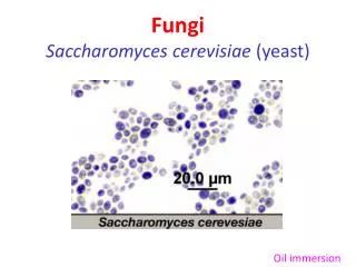
Virus of Yeast Virus padaragiSaccharomycescerevisiae
Virus of Yeast Saccharomyces cerevisiae killer virus M1 Saccharomyces cerevisiae disebut “killer” karena membawa ds-RNA virus yang menyebabkan mereka dapat mensekresi sejumlah toksin yang bersifat letal untuk sel tertentu Virus M1 (kategori M spesies) merupakan jenis double stranded RNA (ds-RNA) virus pada S.cerevisiae yang berukuran1.7-2.1 kbp (lebih kecil dari L spesies yang berukuran 4.5) Termasuk cytoplasmic viruslike particles (VLPs). Saccharomyces cerevisiae killer virus M1 mengkode toksin K1 dan K28 yang spesifik untuk mekanisme imunitas
Virus of Yeast Saccharomyces cerevisiae killer virus M1 Produk protein inisial dari proses translasi M ds-RNA disebut prepotoksin. Target prepotoksin adalah secretory pathway dari yeast. Prepotoksin diproses untuk menghasilkan α/β dimer yang bertindak sebagai bentuk aktif dari toksin Mekanisme Toksin K1: K1 berikatan dengan β-1,6-D-glucan receptor pada dinding sel target masuk ke dalam sel berikatan dengan reseptor plasma membran Kre1p Membentuk kation-selektif channel ion pada membran yang bersifat letal pada sel
Virus of Yeast Saccharomyces cerevisiae killer virus M1 Mekanisme toksin K28: K28 menggunakan reseptor α-1,6-mannoprotein untuk masuk ke dalam sel Toksin prekursor dapat diimpor ke retikulum enoplasma setelah diproses di RE, K28 pindah ke sitoplasma dan menghentikan sintesis DNA pada nukleus, meransang terjadinya apoptosis
Virus of Yeast Overview of the Killer Yeast • Killer yeast = yeast (Saccharhomyces cereviseae) which carry a double stranded RNA virus. • These viruses the yeast to secrete a number of toxic proteins which are lethal to receptive cells. • These yeast cell are immune to the toxic effects of the protein due to an intrinsic immunity. • Killer yeast strain can be a problem in commercial processing kill desirable strains.
Saccharomyces cerevisiae virus L-A Virus classification: Group: Group III (dsRNA) Family: TotiviridaeGenus: Totiviru The Taxonomy of the Virus
The virus discussed above = Saccharomyces cereviseae virus L-A. Icosahedral dsRNA virus. It has a single 4.6 kb genomic segments encodes is major coat protein: Gag (76 kDa) and a Gag–Pol fusion protein (180 kDa) and several satellite double-stranded RNA sequences = M dsRNAs. The genomic segment encodes: a. the viral coat protein b. protein which replicates the viral genomes The M dsRNAs encodes: a. secreted protein toxin (the Killer Toxin). b. immunity to the toxin. About the Virus
The initial protein product from the translation of the M dsRNA = propotoxin. The toxin is targeted to the yeast secretory pathway. The toxin is processed and cleaved to produce an alpha/beta dimer active form of the toxin. The most studied variant toxins in S.cereviseae: a. K1 : binds to the β-1,6-D-glucan receptor on the target cell wall, moves inside, and then binds to the plasma membrane receptor Kre1p. It forms a cation-selective ion channel in the membrane lethal to the cell. More about the Killer Toxins
b. K28 : uses the α-1,6-mannoprotein receptor to enter the cell, and utilizes the secretory pathway in reverse by displaying the endoplasmic reticulum HDEL signal. From the ER, K28 moves into the cytoplasm and shuts down DNA synthesis in the nucleus apoptosis. What’s Next About the Toxin?
Several experiments have made use of this to reliably indentify strains Morace, Archibusacci, Sestito and Polonelli (1984) used the toxins produced by 25 species of yeasts to differentiate between 112 pathogenic strains, based on their sensitivity to each toxin. Used to control undesirable yeast Polonelli et al. (1994) used a killer yeast to vaccinate against C. albicans in rats. Used as an antifungal agents Several experiments suggest that antibodies that mimic the biological activity of killer toxins have application as antifungal agents. The Uses of the Toxins
5S RNA and tRNA-like Molecules Are Associated with Killer Virus Killer virus ialah virus yang diturunkan (inherited) di sitoplasma pada sel-sel yeast (ragi). Sel-sel yeast yang menjadi inang virus (killers) mensekresi suatu protein toxin yang lethal (mematikan) terhadap sel-sel yang sensitif/tidak mengandung virus Genom killer virus terdiri dari 2 segmen dsRNA yang terkapsidasi dalam sitoplasmik virion pada sel-sel yang terinfeksi.
Informasi genetik untuk toxin dan resistensi dikode pada M dsRNA (1830 bp) yang mendandung 200 bp AU-rich region. Segmen dsRNA lain, disebut LA (4980 bp) mengkode sebagian besar protein kapsid (M dan LA terkapsidasi). Molekul dsRNA lain yang disebut LB dan LC terdapat di beberapa sitoplasma, baik killer maupun non-killer yeast.
dsRNA virus pada S. cerevisiae terdiri dari 4.5 kb spesies L dan 1.7-2.1 kb spesies M, keduanya ditemukan pada partikel menyerupai virus di sitoplasma (viruslike particles/ VLPs). Spesies L mengkode protein kapsidnya sendiri, dan LA mengkode capsid-polimerase fusion protein (cap-pol) yang diasumsikan menghasilkan VLPs dengan fungsi-fungsi replikase-transkriptase nya. M1 dan M2dsRNAs mengkode K1 dan K2 toxin serta fungsi dari replikasi.
This virus prevent growht of sensitive wild yeast srains • Caused contamination of alcoholic fermentation. High volatile activity, H2S production, off-falvors caused by fusel oil,acetaldehyd, lactic acid
Contoh ScV-L • Simple double-stranded RNA • -4,8 kbp RNA (L) encapsidated in isometric • Have two desaperately dsRNAs, • Larger is 4,9 kbp encodes the major capsid polypeptida • Smaller dsRNA (M) is 1,9 kbp encodes a secreted polypeptide toxin (killer factory)
Apa itu Parasit? • Parasitadalahorganisme yang hiduppadainanguntukmemperolehnutrisitanpamemberikankeuntunganpadainangtersebut. Bahkancenderungmerugikanpadainang. • Beberapacontohparasitdiantaranyaadalah protozoa, yeast, atauorganismemultisellularlainnyaseperti fungi ataucacing.
Protozoa • Protozoa merupakan hewan bersel tunggal, berinti banyak dan tidak memiliki dinding sel. Ukurannya antara 3-1000 mikron dan merupakan organisme mikroskopis bersifat heterotrof. • Beberapa jenis protozoa bersifat parasit dan menyebabkan penyakit pada manusia dan hewan ternak.
Penemuan Virus pada Protozoa • Virus atau partikel seperti virus telah terobservasi pada beberapa protozoa, yaitu Leishmania, Entamoeba histolytica, Acanthamoeba sp., Naegleria, Plasmodium vivax, P. berghei, Paramecium aurelia, Carchesilun polypinum dan 19notocoma sabellarum. • Pada strain tertentu Paramecium aurelia ditemukan adanya fenomena ‘pembunuh’ (killer)yang berkaitan dengan keberadaan sejumlah partikel yang disebut sebagai kappa di dalam sitoplasmanya.
Kappa • Status biologis kappa dan partikel terkait, telah lama menjadi subjek investigasi intensif dan memunculkan banyak spekulasi. • Kappa telah diamati sebagai elemen genetika endegenous, virus, rickettsia, bakteri atau alga degenerasi. Bukti yang mendukung bermacam sudut pandang sangat sedikit dan kebanyakan tidak dapat dibuktikan. • Sonneborn (1938) menyebut partikel infeksi, innterselular parasit, menjadi manifestasi sebuah level organisasi protoseluler,yang akan menentukan perbedaannya dari tipe bakteri.
Membedakannya.. • Ukuran, sensitivitas pada antibotik, reproduksi dengan pembelahan transverse dan keberadaan DNA dan RNA pada partikel yang sama akan menjadikan mereka seperti rickettsia atau bakteri. • Sebaliknya, ketiadaan atau sedikitnya enzim dan kegagalan pertumbuhan ekstraseluler akan membuat mereka seperti virus. Konsentrasi DNA dan RNA pada Kappa dan partikel lambda sangat mirip dan tidak dapat membedakan partikel ini dari bakteri.
Keberadaan kappa bergantung kepada gen kromosomal dominan K. Beberapa peneliti, seperti T.M. Sonneborn, mengamati bahwa sel P. aurelia yang mengandung partikel-partikel kappa akan menghasilkan senyawa beracun yang dapat mematikan strain-strain protozoa lainnya yang ada di sekitarnya. Senyawa beracun ini selanjutnya disebut sebagai paramesin, sedangkan partikel-partikel kappa ternyata merupakan bakteri simbion yang kemudian dikenal dengan nama Caedobacter taeniospiralis, yang artinya bakteri pembunuh berbentuk pita spiral.
00.075.0.03.001. Leishmania brasiliensis virus 1-1 Name, Synonyms and Lineage Synonym(s): Leishmania RNA virus-1. ICTV approved acronym: LRV1- 1. Virus is the type species of the genus Leishmaniavirus; family 00.075.Totiviridae. ICTVdB Virus Code: 00.075.0.03.001. Virus accession number: 75003001. Obsolete virus code: 75.0.3.0.001; superceded accession number: 75030001. NCBI Taxon Identifier NCBI Taxonomy ID: 58103.
Virion Properties Leishmania brasiliensis virus 1-1 • Morphology Virions consist of a capsid. Virus capsid is not enveloped, round with icosahedral symmetry. The isometric capsid has a diameter of 33 nm. • Nucleic Acid The genome is monopartite, only one particle size is recovered of linear, double-stranded RNA. The complete genome is 5284 nucleotides long, is fully sequenced. Sequence has the accession number [M92355]. The 5'-end of the genome does not have cap. GenBank records fornucleotide sequences; complete genome sequences. • Proteins The viral genome encodes structural proteins and non-structural proteins. • Lipids Lipids are not reported.
Biological Properties Leishmania brasiliensis virus 1-1 Natural Host • DomainViral hosts belong to the Domain Eucarya. • Domain EucaryaKingdom Protoctistae.
Giardia lamblia Giardia lamblia adalah protozoa penyebab giardiasis, mengganggu pencernaan, dengan berdiam di usus. Menginfeksi dengan membentuk cyst pada saluran pencernaan dan menyebabkan diare.
00.075.0.02.001. Giardia lamblia virus Name, Synonyms and Lineage ICTV approved acronym: GLV. Virus is the type of the genus 00.075.0.02.Giardiavirus; of the family 00.075.Totiviridae. CTVdB Virus Code: 00.075.0.02.001. Virus accession number: 75002001. Obsolete virus code: 75.0.2.0.001; superceded accession number: 75020001. NCBI Taxon Identifier NCBI Taxonomy ID: 29255.
Virion PropertiesGiardia lamblia virus • Morphology Virions consist of a capsid. Virus capsid is not enveloped, round with icosahedral symmetry. The isometric capsid has a diameter of 36 nm. • Nucleic Acid The genome is monopartite, only one particle size is recovered of linear, double-stranded RNA. The complete genome is 7000 nucleotides long, is fully sequenced. Sequence has the accession number [L13218]. The 5'-end of the genome does not have cap. GenBank records fornucleotide sequences; complete genome sequences. • Proteins The viral genome encodes structural proteins and non-structural proteins. • Lipids Lipids are not reported.