Download
1 / 23
230 likes | 312 Views
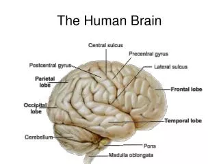
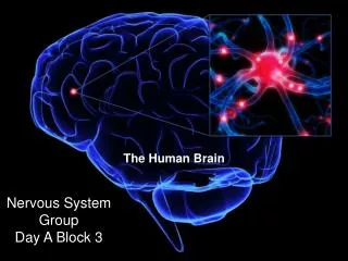
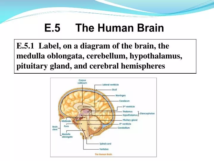
E.5 The Human Brain. Cerebral Hemispheres. Medulla oblongata • The most inferior part of the brain stem • Controls autonomic and homeostatic activities such as heart rate, blood vessel diameter, respiratory rate, vomiting, coughing, swallowing, and digestion

E N D
Medulla oblongata • The most inferior part of the brain stem • Controls autonomic and homeostatic activities such as heart rate, blood vessel diameter, respiratory rate, vomiting, coughing, swallowing, and digestion • It is also a conduction pathway between higher brain centers and the spinal cord
Cerebellum •Regulates balance and equilibrium as well as posture • It also coordinates skeletal muscle movements Hypothalamus • The chief integration center for the ANS • Regulates body temperature, food intake, water balance, thirst, and biological rhythms and drives • Regulates hormonal output of anterior pituitary gland as well as being an endocrine gland itself by producing oxytocin and ADH • Part of the limbic system which is involved with emotional responses and memory processing
Pituitary Gland • A neuroendocrine gland located beneath and attached to the hypothalamus by a stalk (infundibulum) • It regulates the gonads, thyroid gland, adrenal gland, lactation, and water balance through the production and release of pituitary hormones Cerebral Hemispheres • Form the superior part of the brain and account for 83% of the total brain mass • Contain grey matter (cerebral cortex) that localizes and interprets sensory inputs, controls voluntary muscle activity • Also function in intellectual and emotional processing • Contain basal ganglia that are important in the initiation of skeletal muscle movements
• It has recently been discovered that magnetic resonance imaging can be used to map changes in brain hemodynamics that correspond to mental operations, extending traditional anatomical imaging to include maps of human brain function • Preliminary investigations of human brain mapping with this procedure has yielded insights into the functional organization of various sensory, motor, and language systems
• Functional MRI is based on the increase in blood flow to the local vasculature that accompanies neural activity in the brain Increased emotions and arousal
• The main advantages to this technique include 1). The signal does not require injections of radioactive isotopes, 2). Total scan time required can be very short (1.5 -2.0 min. per run), 3). The resolution of the functional image is good • Animal experiments have been used to test the effect of exercise on brain function (Neurobiology of Disease, Vol. 13, Issue 1, June 2003, pp 1-14) • It was found that exercise leads to increased serum calcium levels • The calcium is transported to the brain and enhances the synthesis of dopamine
• The increased dopamine levels regulate various brain functions • Low dopamine levels were found in epileptic mice and spontaneously hypertensive rats and improvement was noted following exercise, or through intracerebroventricular administration of calcium chloride • The findings indicated that exercise or convulsions affect brain function through calcium-dependent dopamine synthesis leading to the possibility that some symptoms of Parkinson’s disease or senile dementia might be improved by exercise
• Lesions in specific areas of the brain change behavior in specific ways • Studies have been done to correlate these behavioral changes with the site of the lesions and provided information that can be used to predict from a given behavioral disturbance the site of the lesions, and vice versa • This information can be used clinically (Clinical Neuropsychology)
• When under stress, sympathetic stimulation of the heart causes increased contractility (strength) through the release of norepinephrine which causes the pacemaker to fire more rapidly, and by enhancing the entry of calcium ions into the heart cells; the result is increased heart rate • The parasympathetic division opposes sympathetic effects and reduces heart rate when a stressful situation has passed
• The iris or the eye has as part of its structure, two layers of smooth muscle fibers which can change the diameter of the central opening (pupil) •Both muscle layers are controlled by the ANS • The dilator muscles of the iris are stimulated by the sympathetic division, thus dilating eye pupils in response to dim light • The parasympathetic division stimulates contractor muscles which constrict eye pupils in response to bright light
• Although the digestive system responds to many different types of controls, i.e. hormones, pH, etc., the parasympathetic nervous system is crucial to its normal functioning • The parasympathetic division increases peristalsis and the amount of secretion by digestive organs as well as relaxing sphincters to allow movement of foodstuffs along the tract • The sympathetic division decreases activity of glands and muscles of the digestive system and constricts sphincters
• The pupil reflex allows for light and dark adaptation whereby reflexive changes occur in pupil size • Bright light shining in one or both eyes causes both pupils to constrict • These pupillary reflexes are mediated by the pretectal nucleus (branch from the optic nerve) of the midbrain and by parasympathetic fibers • In dim light, the pupils dilate, allowing more light to enter the eye interior
• Pupils are examined for size, shape, and equality • Pupillary reflex is tested with a penlight (pupils should constrict when illuminated); this reflex is important since the retina can be damaged by too much light • The midbrain of the brain stem contains the superior and inferior colliculiwhich are visual and auditory reflex centers
•Death is defined as the absence of brain activity; spontaneous brain waves are present even during unconsciousness and coma, therefore, the pupillary reflex should still be present •Failure of the pupils to respond to light is indicative of damage to the brain stem; pupils remain fixed and dilated due to the absence of brain activity •Confirmation can be accomplished by doing an EEG which is a test used for the diagnosis and localization of many types of brain lesions, such as tumors, abscesses, etc. • Interference with cerebral cortical functions is suggested by brain waves and clinical evidence of brain death results in a “flat” EEG
• Clinically, pain is classified as somatic (arising from skin, muscles, or joints) or visceral (arising from receptors in the organs of the thorax and abdominal cavity) • Somatic pain may be superficial or deep while visceral pain is deep • Both superficial somatic pain fibers and deep somatic/visceral pain fibers synapse with interneurons in the spinal cord which then transmit pain impulses to the CNS along sensory neurons
• This transmission causes the release of glutamate and substance P,* the pain neurotransmitter, into the synaptic cleft resulting in depolarization of the postsynaptic membrane • The axons of these interneurons are believed to ascend to the thalamus and from the thalamus, impulses are relayed to the cerebral cortex for interpretation • For some time, it has been known natural opiates, endorphins, are released in the brain and spinal cord when we are in pain and act to reduce its perception when under certain stressful conditions
• Endorphins are neuromodulators (make adjustments) and appear to inhibit pain perception by causing blood vessels to dilate; blood flow around joints and muscles increases, thus augmenting nutrient and oxygen delivery to problem areas (runner’s high); they also are carried to the brain and bind to pain receptors blocking the release of the neurotransmitter that is used to transmit pain signals to the brain (substance P) *Substance P – a neuropeptide; an important mediator of pain signals - associated with inflammatory processes and pain perception in CNS