Download
1 / 68
680 likes | 1.03k Views
Does Chlamydia pneumoniae stimulate the inflammatory process of coronary atherosclerosis?. David E. Cox, M.D. Resident Grand Rounds January 9th, 2001. Overview. Introduction. Atherosclerosis as an inflammatory process. Infectious agents and atherosclerosis.

E N D
Does Chlamydia pneumoniae stimulate the inflammatory process of coronary atherosclerosis? David E. Cox, M.D. Resident Grand Rounds January 9th, 2001
Overview. Introduction. Atherosclerosis as an inflammatory process. Infectious agents and atherosclerosis. The case for Chlamydia pneumoniae. Seroepidemiologic studies. Biologic plausibility. Histologic studies. Animal models. Retrospective analysis of antibiotic use and risk of MI. Existing and future randomized controlled trials.
INTRODUCTION. Atherosclerosis. • Appropriately described as an inflammatory process. • Initial insult occurs in the endothelium. • Endothelial denudation or alteration of normal endothelial homeostatic mechanisms. • Deleterious factors include hypertension, elevated and modified LDL, free radicals from cigarette smoking, or diabetes mellitus. • Less well-recognized risk factors include elevated homocysteine, Lp(a), genetic factors, and possibly infectious agents.
Atherosclerosis (cont.) • Trapping of lipoproteins in the vessel wall. • Earliest of events. • Abnormal expression of proteoglycans in the endothelial cell wall that have increased binding affinity for lipoproteins. • Altered endothelial homeostatic mechanisms. • Increased expression of platelet and leukocyte adhesion molecules. • Increased cellular permeability. • Balance shift from anticoagulant to procoagulant properties. • Liberation of growth factors, vasoactive molecules, and leukocyte chemoattractants (monocyte-chemotactic protein)
Atherosclerosis (cont.) • Smooth muscle cells. • Proliferate and migrate into the lesion • Lose contractile phenotype, express growth factors, cytokines, etc. • Macrophages. • Monocytes and T lymphocytes are drawn to the lesion. • Monocytes enter the vessel wall and become tissue macrophages. • Macrophages respond to oxLDL by expressing scavenger receptors. • Scavenger receptors bind more oxLDL, which macrophages internalize and transform into foam cells. • Foam cells secrete more growth factors, cytokines, andchemoattractants that recruits more LDL and inflammatory cells into the lesion.
Atherosclerosis (cont.) • The advanced lesion. • Macrophages, T lymphocytes, and transformed smooth muscle cells are activated to secrete hydrolytic enzymes, cytokines, chemokines, etc. • Eventually the lesion advances to a necrotic core of lipids and cellular debris covered by a fibrous cap. • Remodelling of the vessel. • As the lesion develops, compensatory mechanisms by the vessel limit the impingement of blood flow. • Eventually compensatory mechanisms of vessel dilatation are overcome and the lesion becomes stenotic, impairing flow.
Atherosclerosis (cont.) • Stable lesions. • Uniformly dense fibrotic caps. • Usually the more stenotic ( 75% occlusion). • Less prone to acute plaque rupture. • Unstable lesions. • Usually the nonobstructive lesions. • More prone to acute plaque rupture. • Plaque shoulders are the rupture points. • Metalloproteinases liberated by macrophages may thin the fibrous plaques in these areas. • Leads to thrombus formation over the plaque. • Presents in the spectrum of acute coronary syndromes.
Effect of infectious organisms on atherosclerosis. May upregulate the inflammatory process. • Influx of inflammatory cells. • Stimulate the release of immunomodulators. Potential clinical outcome. • Plaque progression to vessel stenosis. • Acute plaque rupture.
Chronic diseases associated with infectious agents. Peptic ulcer disease H. pylori Dementia T. pallidum Guillain-Barre Campylobacter Glomerulonephritis Streptococcus Colon cancer Strep. bovis
Infectious agents purported to have pro-atherosclerotic effects. Enterovirus Adenovirus HSV-1 HSV-2 CMV H. pylori Chlamydia pneumoniae
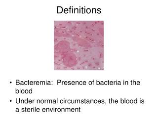
Seroepidemiologic studies of H. pylori and atherosclerotic disease.
Seroepidemiologic studies of CMV and atherosclerotic disease.
Seroepidemiologic studies of C. pneumoniae and atherosclerosis.
Clinical Case. Mr. B is a 65 m with h/o CAD, s/p MI 1995 with multiple percutaneous interventions presents to the outpatient clinic with 3 day history of cough productive of purulent sputum, malaise, and low grade fever. PMHx CAD, MI 1995 DM HTN Dyslipidemia PAD: ruptured AAA repaired ‘95 h/o bilateral iliac bypass
Clinical Case (cont.) Exam T99.2 BP150/85 P75 R18 PO2 96%RA NAD, O/P clear, PERRLA/EOMI, no JVD, no sinus tenderness; bilat. rhonchi, no rales; RRR no mgr; no edema, 2+ pulses. CXR: NACPD, Labs: CBC, electrolytes nl Meds: Lopressor, Accupril, Lasix, Lipitor, ECASA, Paxil. Dx: Acute bronchitis, probably viral etiology. Tx: Antitussives, decongestants.
Clinical Case (cont.) QUESTION: Chlamydia pneumoniae has emerged as a causative agent of acute bronchitis, and may induce progression of atherosclerosis; would a course of antibiotics possibly reduce this patient’s risk of an adverse cardiac outcome?
Life cycle of Chlamydia pneumoniae. • Gram-negative, obligate intracellular organism. • Life cycle of the organism is conducive to chronic infection. • Two forms: elementary bodies and reticulate bodies. • Elementary bodies. • metabolically inactive, although infectious • unaffected by antibiotics • Reticulate bodies. • metabolically active • exclusively intracellular form • affected by antibiotics: macrolides, tetracycline, and quinolones.
R E
Clinical implications of C. pneumoniae. Clinical manifestations. • Sinusitis, bronchitis, pneumonia. Prevalence of infection is high. • Over 50% persons have first infection by age 20. • Reinfection is common. • IgG antibodies are usually lost by 3-5 years post infection. • The prevalence of serum IgG antibodies continues to rise with age. • 80% in men by age 80. • 70% in women by age 80. • This data suggests either frequent reinfection or recrudescence of prior infection.
The case for Chlamydia pneumoniae. Seroepidemiologic studies. Biologic plausibility. Histologic studies. Animal models. Retrospective analysis of antibiotic use and risk of MI. Existing and future randomized controlled trials.
Seroepidemiology: Saikku et al. Evaluation of three groups in Helsinki b/t ‘83 and ‘85. 40 consecutive men admitted for AMI 30 consecutive men admitted for cardiac catheterization • secondary to chronic angina • all with prior evidence of coronary artery disease, 19 with prior MI • referred to as the chronic coronary heart disease (CCHD) group 41 randomly selected aged-matched male controls • no evidence of CAD as per negative history and EKG • selected within 1-3 weeks of admission
Saikku et al. (cont.) Serologic samples • IgG/IgA antibodies to C. pneumoniae measured upon admission • Antibody measured using microimmunofluorescence • Antibody titers of > 1/32 considered positive
Seroepidemiologic studies of C. pneumoniae and atherosclerosis. 17 serologic studies after Saikku et al. • Inherent weaknesses: • different populations. • different criteria for adverse cardiovascular endpoints. • adjusted for confounders to different degrees. • Post-hoc analysis: • various cut-off points used to define titer seropositivity. • sometimes not chosen until data was evaluated. • Microimmunofluorescence: • interpreted by expert microscopists. • subject to bias
Seroepidemiologic evidence:Key Points. 1. Initial observations revealed a positive association of acute MI and chronic coronary heart disease with the presence of anti-Cp antibodies. 2. The increased antibody titers tended to be more strongly associated with acute MI and coronary heart disease. 3. Subsequent serologic studies favor an association between various surrogate endpoints of coronary heart disease and elevated anti-Cp antibody titers. 4. This evidence sets the foundation for further investigation to establish a causal role for Cp and coronary heart disease.
“Hello, PAL line? Yeah, this is Mt. Airy; hey, we’ve got a 24 year old female with chest pain and the ‘sniffles.’ We’re going to transfer her to Dr. Kirkman for intensive Z-pak and IV vitamin E therapy.”
The case for Chlamydia pneumoniae. Seroepidemiologic studies. Biologic plausibility. Histologic studies. Animal models. Retrospective analysis of antibiotic use and risk of MI. Existing and future randomized controlled trials.
Biologic Plausibility. Are there pathophysiologic mechanisms? • How does C. pneumoniae get into the lesion? • How might C. pneumoniae participate in the process of atherogenesis and/or plaque destabilization?
How does C. pneumoniaeget into the lesion? • Dissemination of organism from sinopulmonary infection. • Rosenfield et al. • respiratory inoculation mice peritoneal Cp-infected M. • Godzik et al. • all cell types of the vessel wall can be infected (endothelial and smooth muscle cells, and fibroblasts) in vitro. • Kaukoranta et al. • Cp-infected monocytes have also been shown to transfer infection to endothelial cells in vitro. • Cp-infected endothelial cells upregulate expression of leukocyte adhesion molecules.
How might C. pneumoniae participate in atherogenesis and/or plaque destabilization? • C. trachomatis and psittacii can stimulate granulocytes to release oxidants (cellular respiratory burst). • Cp-infected M • Cp-induced release of oxidants from M could serve to increase formation of oxLDL. • may also express more scavenger receptors and be more prone to developing into foam cells. • may be upregulated in the production of immunomodulators. • M presentation of Cp antigens may stimulate the influx of T lymphocytes.
Biologic Plausibility:Key Points. 1. Cp has in vitro capability to infect various cells that comprise the atherosclerotic lesion and the vessel wall. 2. Cp infection of the monocyte cell line induces proatherogenic changes in vitro: • increased formation of oxLDL • influx of T lymphocytes • stimulation of cellular products foam cell formation • increased leukocyte adherence to endothelium 3. This adds some strength to a causal association, but more evidence is required to assess clinical significance.
The case for Chlamydia pneumoniae. Seroepidemiologic studies. Biologic plausibility. Histologic studies. Animal models. Retrospective analysis of antibiotic use and risk of MI. Existing and future randomized controlled trials.
Histologic Studies. CASE REPORT: A 39 y South African Male. • Died of an acute MI. • No risk factors for CAD except mild HTN. • Serology prior to death confirmed seropositivity to Cp. • Autopsy: LAD ruptured plaque. • Pathology: Cp detected by PCR, EM, and ICC in the smooth muscle cells and foam cells of the plaque. • Organism was also detected by these methods in the anterior wall myocardium, suggesting Cp was released from the atheroma in the LAD and disseminated into the area of infarct.
Histologic studies (cont.). 30 studies identified C. pneumoniae in atheromatous tissue. • PCR, Electron microscopy (EM), immunocytochemistry (ICC). • Culture of live organism from plaques has been rare. Kuo et al.: Detection of C. pneumoniae by PCR and ICC. • 50% of atheromatous vessels • 1% of normal vessels Pathologic Determinants of Atherosclerosis in Youth (PDAY) • Cp detected by PCR or ICC • 6/7 (86%) atherosclerotic plaques. • 2/11 (18%) lesions with intimal thickening. • 0/31 (0%) normal arteries.
Histologic studies (cont.). “Innocent bystander” hypothesis • C. pneumoniae exists in monocytes in its active form. • Monocytes taken up into atheroma • C. pneumoniae may just “go along for the ride.”
Histologic studies:Key Points. 1. Cp antigens and Cp DNA has been detected within atherosclerotic plaques to a much greater degree than normal tissues. 2. Culture of live organism from plaques has been rare. 3. Cp may be “innocent bystanders” in the plaque. 4. This evidence adds to the concept of biologic plausibility, but still begs for more clinical correlation.
The case for Chlamydia pneumoniae. Seroepidemiologic studies. Biologic plausibility. Histologic studies. Animal models. Retrospective analysis of antibiotic use and risk of MI. Existing and future randomized controlled trials.
Animal Models. Moazed et al. • ApoE-deficient mice spontaneous atherosclerotic lesions. • Infected with C. pneumoniae via intranasal innoculation. • Mice euthanized, C. pneumoniae was demonstrated in atheromatous plaques in the aorta by PCR and ICC.
Animal Models (cont.). Muhlestein et al. • 30 New Zealand white rabbits • 20 received intranasal innoculation of C. pneumoniae. • 10 received saline. • 3 week intervals for 3 cycles. • All rabbits fed cholesterol-rich diet. • After 3 innoculations • 10 infected rabbits rec’d 7 wk. course azithromycin, 10 rec’d placebo. • 5 non-infected rabbits received azithromycin, 5 received placebo. • 3 months after the final innoculation, the rabbits were euthanized.
Animal Models (cont.). Muhlestein et al. (cont.) • Major endpoints. • maximal intimal thickness (MIT) of thoracic aorta. • plaque area index (PAI) = MIT x the % luminal circumference. • Results. • There was no difference b/t control groups that rec’d abx v. placebo. • There was no difference b/t infected/treated and control groups. • There was a significant difference b/t infected/untreated and both control and infected/treated groups. • Infected/untreated MIT = 0.55 ± 0.15 mm. • Infected/treated MIT = 0.16 ± 0.06 mm. • Control group MIT = 0.20 ± 0.03 mm.
Animal Models:Key Points. 1. Mouse models which develop spontaneous atherosclerotic lesions have been infected with Cp and shown to localize organism to aortic atheromas. 2. A few studies have shown Cp-induced atherogenesis in rabbit aorta can be arrested using macrolide antibiotics. 3. This provides more evidence regarding the potential effects of Cp’s contribution to atherosclerosis in vivo.
The case for Chlamydia pneumoniae. Seroepidemiologic studies. Biologic plausibility. Histologic studies. Animal models. Retrospective analysis of antibiotic use and risk of MI. Existing and future randomized controlled trials.
Prior Antibiotic Use and Incidence of First-Time Acute Myocardial Infarction. JAMA 1999. Meier et al. • Data extracted from UK General Practice Research Database • 3315 pts. with first-time MI matched to 13,139 controls w/o prior MI. • Exclusion: h/o ischemic heart disease, CHF, arrhythmia dyslipidemia hypertension diabetes mellitus • Previous antibiotic use over 3 years was reviewed. • Odds ratios were adjusted for tobacco use and BMI.
Prior Antibiotic Use and Incidence of First-Time Acute Myocardial Infarction.
Prior Antibiotic Use and Incidence of First-Time Acute Myocardial Infarction. Tetracyclines • good activity against C. pneumoniae. Quinolones • marginal activity against C. pneumoniae. • effect here in small numbers (8/3315 cases and 62/13,139 controls). Beta-lactams, cephalosporins, and sulfonamides • Poor activity against C. pneumoniae. Macrolides • typically have good coverage for C. pneumoniae. • Erythromycin most commonly used in these patients.