Download
1 / 43
530 likes | 1.28k Views
Honors Biology Microscopes. Important tool for all biologists. Honors Bio: Microscopes. Use light or electrons to magnify Enable us to see the shape and structure of very small objects Cells and cell parts Tissues Molecules (only with electron microscopes)

E N D
Honors BiologyMicroscopes Important tool for all biologists
Honors Bio: Microscopes Use light or electrons to magnify Enable us to see the shape and structure of very small objects • Cells and cell parts • Tissues • Molecules (only with electron microscopes) • Small and microscopic organisms
Value of MagnificationReal size Magnified 400 X Cell walls cytoplasm Elodea canadensis Pond weed chloroplasts central vacuole
Magnification Magnification = object size ~ image size Total magnification = ocular lens X objective lens chloroplasts flagellum nucleus food vacuole Euglena, a one-celled organism 1000X
Resolution or Resolving Power Resolution = sharpness, clarity of focused image • “Ability to show two close points as separate” • Depends on shape and perfection of lenses • Human eye can see objects as small as 0.2 mm • A light microscope can resolve objects as small as 0.2 m high resolution lens lower resolution lens
Depth of Field • Thickness or layer in focus • Higher magnification thinner layer
Light MicroscopesSend LIGHT through a thin specimen an early microscope binocular light microscope
Light Microscopes (LM) • Light waves pass through a thin specimen • Lenses bend light to magnify image • Simple microscope – one lens • Compound microscope – two lenses • Magnifies image twice
Leeuwenhoek’s Microscope • Anton von Leeuwenhoek, 1600s • First powerful scope with high resolution • Single lens • Magnify ~ 300 X
Eyepiece Ocular lens LE 4-1a Objective lens Specimen Condenser lens Light source BINOCULAR MICROSCOPE – has ocular lens for each eye
Epithelial cell Photosynthetic cells Chloroplast (dots inside cell) Stoma (leaf opening) Leaf cross-section(LM)
Advantages of light microscopes • Can magnify up to 2000 times • Shows shape and structure of cells and tiny organisms • Specimens can be alive Disadvantages • Specimens must be thin enough for light to pass through • Image appears inverted and backwards • Often need stain to see image
Cheek cells with stain Light microscope LM “dark field” Common stains: methylene blue, Lugol’s iodine “Vital stains” - stain without killing cells
Phase-Contrast Microscope“Differential Interference Microscope” Increases contrast between tissue densities – don’t need stain; good for living organisms Cheek cells without stain
Phase-Contrast Microscope cheek cells –unstained Compound Microscope cheek cells – stained nucleus cytoplasm cell membrane nucleus cytoplasm cell membrane
Amoeba, one-celled organism preserved, stained alive, moving Compound scope Phase-Contrast scope
Stereomicroscope“Dissecting microscope” Has ocular lens and objective lens for each eye Stereoscopic vision, 3-D Image NOT inverted Magnifies 10-50X
Advantages of stereoscopes • Image NOT inverted or backwards • Makes manipulation easy • Specimens can be solid, living • Disadvantage: magnifies up to ~50 X
Stereomicroscope – whole specimens chick embryo soil worm
Fluorescent Microscopy • Uses lasers on thin slices; confocal scope • Fluorescent dyes show different molecules Cancer cells tagged with 3 fluorescent dyes shows cell microtubules (blue), microfilaments (yellow), DNA (green)
Fluorescent – shows different cell parts as different colors • Details in a single layer Fruit fly embryo – developmental layers Green – microtubules in cytoplasm Red -DNA • http://www.microscopyu.com/tutorials/java/virtual/confocal/index.html
Confocal Microscopy Specialized Cells in the Ear E. Coli bacteria
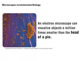
Electron Microscope • Uses electrons instead of light • Magnets focus the beam • Image shows on monitor • Magnify up to 1 million times • Show cell details, interior • - “ultrastructure • Invented 1930’s • Nobel for Ruska 1986
Electron Microscope • How does it work? • Specimen is coated with a metal film • Electron beam hits metal, ejects electrons from metal atoms • These electrons make the image
Advantagesof electron microscopy • Electron are much smaller than the wavelength of light – show things that light cannot show • Very high magnification – up to 1,000,000X • Very high resolution - up to 1 nanometer • DISADVANTAGE – specimen must be dead, dried, coated, in vacuum chamber
Scanning Electron MicroscopeSEM • Electron beam skims across specimen surface • Shows tiny surface structures in great detail • Magnifies up to 50,000 times • DISADVANTAGE: shows surface, but not interior
Compare LM and SEM Blood cells (LM) Blood cells (SEM)
SEM micrographs Euglena(protist) SEM Ant head, SEM
Scanning Electron Microscope (SEM) shows surface details Electrons scan across surface of specimen
SEM of DNA Image made with special scanning “tunneling” microscope
Transmission Electron Microscope (TEM) shows inside cells • Electrons pass through thin specimen • Shows great detail of internal structure • Magnifies up to 1,000,000 times!! Rough ER Mitochondria Nucleus
Transmission Electron Microscope Bacterium dividing Muscle fibers Phage virus Liver cells Cilia and basal bodies Chloroplast
Comparing microscopes Euglena SEM Euglena LM Euglena TEM
Which type of microscope produced these micrographs? Amoeba, preserved and stained Vacuole inside a cell Amoeba, alive and unstained
Which type of microscope made these micrographs? Female and male fruit fly Closterium -Unicellular green alga
Name the microscope Leaf cross-section 400X chloroplast 5,000 X
Name the microscope Iridescent beetle Eye of a housefly