Download
1 / 33
330 likes | 755 Views
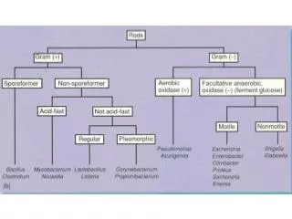
Corynebacterium & Bacillus. - Microscopic appearance - Colonial morphology. Corynebacterium diphtheriae. - Nasal, nasopharyngeal and tonsillar diphtheria. Corynebacterium diphtheriae. - Cutaneous diphtheria. Corynebacterium diphtheriae. - Throat, nasopharyngeal swabs. - Skin swab.

E N D
Corynebacterium & Bacillus • -Microscopic appearance • -Colonial morphology
Corynebacteriumdiphtheriae - Nasal, nasopharyngeal and tonsillar diphtheria.
Corynebacteriumdiphtheriae - Cutaneous diphtheria.
Corynebacteriumdiphtheriae - Throat, nasopharyngeal swabs. - Skin swab.
Corynebacteriumdiphtheriae - Gram positive pleomorphic, long, thin, and curved forms can be seen and also short rods and rods enlarged at one end (clubshaped).
Corynebacteriumdiphtheriae - They often appear in clusters, joined at angles like Chinese letters.
Albert Staining of volutin granules • C. diphtheriae often appears beaded due to the presence • of dark staining granules in the rods. • These granules, known as volutin or metachromatic • granules, are energy-storing inorganic polyphosphate • units. In some strains the granules form at the ends of • the rods. • - The granules are most numerous after the organism has • been cultured on a protein-rich medium such as Dorset • egg or Loeffler serum.
Albert Staining of volutin granules 1- Fix the dried smear using alcohol. 2- Cover the smear with the toluidine blue malachite green stain for 3–5 minutes. 3-Wash off the stain with clean water. 4- Tip off all the water.
Albert Staining of volutin granules 5- Cover the smear with Albert’s iodine for 1 minute. Wash off with water. 6- Wipe the back of the slide clean, and place it in a draining rack for the smear to air-dry. 7- Examine the smear microscopically to look for bacteria containing metachromatic granules
Albert Staining of volutin granules Bacteria cells . . . . . . . . . . . . . . . . . . . . . . Pale green Metachromatic granules . . . . . . . . . . . . Green-black
Corynebacteriumdiphtheriae - They often appear in clusters, joined at angles like Chinese letters.
Tinsdal medium:-grey-black raised colonies surrounded by a dark brown area.
Brown colour due to formation of H2S which result from • interaction between cystine and tellurite.
C. diphtheriaegrows rapidly onthese media, producing significant growth in 4–6 hours.
Bacillus anthracis - CutaneousANTHRAX.
Bacillus anthracis - Pulmonary ANTHRAX.
Bacillus anthracis - Enteric ANTHRAX. - Meningoencephalitis.
B. anthracisis a high risk infectious pathogen, therefore handle specimens and infected material with care, wearing protective gloves and face mask, and following recommended safety procedures.
Bacillus anthracis - Fluid aspirated from cutaneous lesions. - Blood for culture. - Sputum. - CSF.
large, 5–8 X 1.5 μm, Gram positive, non-motile bacillus, often appearing joined end to end in chains
In smear from Specimens:- Bacilli are capsulated. The capsular material often appears irregular and fragmented
In smears from aerobic cultures: Bacilli are non-capsulated but contain oval spores (same diameter as the bacilli), giving the organisms a beaded appearance. They occur in chains.
Fixation of smears:- B. anthracisis not killed by heat-fixation. Smearsshould be chemically fixed by immersing the dry smears in a container of potassium permanganate solution for 10–15 minutes.
Bacillus anthracis Blood agar:-large grey-white 2-5 mm in diameter irregular with wavy edges colonies (non or slightly haemolytic).
Bacillus cereus - Food poisoning. - Opportunistic infections in immunocompromised persons (bacteraemia, pnumonia and wound infection).
Bacillus cereus Blood agar:-large grey-white 2-5 mm in diameter irregular with wavy edges haemolytic colonies.
B.cereusrapidly liquefies the gelatin along and out from the line of inoculation.