Download
1 / 40
420 likes | 647 Views
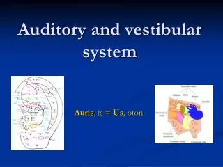
Chapters 9,10 Auditory and Vestibular Systems. Chris Rorden University of South Carolina Norman J. Arnold School of Public Health Department of Communication Sciences and Disorders University of South Carolina. Audition.

E N D
Chapters 9,10 Auditory and Vestibular Systems • Chris Rorden University of South Carolina Norman J. Arnold School of Public Health Department of Communication Sciences and Disorders University of South Carolina
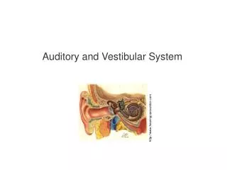
Audition • The ear converts sound energy into patterns of neural firing: transduction • Outer ear: collect and amplify sound, aid in localization • Middle ear: impedance matching • Cochlea: frequency and intensity analysis • Auditory pathway: complex signal processing
Ear structures • Peripheral • Outer ear • Middle ear • Inner ear • Auditory nerve • Central • Brainstem • Midbrain • Cerebral
Anatomy and function • Pinna: the projecting part of the ear lying outside of the head (also called auricle, or just ear lobe) • Reflection of sound in pinna provides spectral cues about elevation of a sound source
Outer Ear – Auditory Canal • External auditory meatus • Provides communication between middle and inner ears by conducting sound to the ear drum • S-shaped tube @ 2.5cm long and @ 7mm wide • Lining of the lateral 1/3rd of canal has cilia and glands • Cerumen (ear wax): protects ear canal from drying out and prevents intrusion of insects
Outer Ear - Ear drum • Tympanic membrane • Separates outer and middle ear • Compliant • Thin, three-layered sheet • Epithelium of EAM: outer layer • Middle layer: fibrous (strong) tissue • Inner layer of middle ear: mucous membrane
Outer Ear - Ear drum • Slightly concave to EAM, cone-shaped • Most depressed and thinnest point is called the umbo • End of the attachment of malleus • ‘Cone of light’ from umbo to periphery reflects light when viewed with otoscope • Slightly oval, taller than wide • Otoscope: if you pull the pinna up and back the tympanic membrane is visible
Role of outer ear • To augment the sound shadow • Ear canal protects delicate parts of middle and inner ear from impact. • To heighten our sensitivity to sounds • Ear canal boosts sounds 15 to 16 dB between 1.5 and 8 kHz (in the area of speech) • This is due to resonance of ear canal • Just like vocal tract this tube amplifies and dampens certain frequencies based on its length and composition
Localization and shadowing • Intensity differences: louder if nearer, less shaded • Inter-aural timing differences • Frequencies influenced by location relative to pinna.
Middle Ear – Eustachian Tube • Establishes communication between middle ear and nasopharynx • ~ 35 to 38 mm long, typically closed • Biological functions: • To permit middle ear pressure to equalize with external air pressure • On the air plane, change in atmospheric pressure but not pressure in middle ear • Yawning or swallowing opens pharyngeal orifice of tube to equalize pressures • To permit drainage of normal and diseased middle ear secretions into the nasopharynx
Middle Ear - Ossicles • 3 of the smallest bones • Malleus (hammer) • Incus (anvil) • Stapes (stirrup) • Ossicular chain: Transmits acoustic energy from tympanic membrane to inner ear • Acts as lever: large weak motion of TM causes small forceful movement of stapes. • Takes force from gas (air) and matches impedance to liquid (inner ear). • Muscles allow movement to be attenuated: Prevents the inner ear from being overwhelmed by excessively strong vibrations
Middle Ear – Ossicles - Malleus Malleus (hammer) 9 mm long Manubrium (handle): attaches to tympanic membrane; pulls the drum medially Caput (head): jointed (quite inflexibly) to Incus
Middle Ear – Ossicles - Incus • The ossicles give the eardrum mechanical advantage via lever action and a reduction in the area of force distribution • Pressure = Force/Area; so less area = more pressure • the resulting vibrations would be much smaller if the sound waves were transmitted directly from the outer ear to the oval window. • The movements of the ossicles is controlled muscles attached to them (the tensor tympani and the stapedius).These muscles can dampen the vibration of the ossicles, in order to protect the inner ear from excessively loud noise and that they give better frequency resolution at higher frequencies by reducing the transmission of low frequencies
Middle Ear – Ossicles - Stapes Head (caput) jointed to incus Anterior and posterior crura (legs) Footplate: joins oval window of inner ear (opening in temporal bone) via annular ligament
Osseous cochlea • Oval window • Connects scala vestibuli and middle ear • Round window • Connects scala tympani and middle ear
Cochlear Structures • Cochleus from 5mo fetus: • Oval window (blue arrow) • Round window (yellow arrow)
Tonotopic • Base • High Freq • Apex • Low Freq.
Travelling wave • Always starts at the base of the cochlea and moves toward the apex • Its amplitude changes as it traverses the length of the cochlea • The position along the basilar membrane at which its amplitude is highest depends on the frequency of the stimulus
Traveling wave • High frequencies have peak influence near base and stapes • Low frequencies travel further, have peak near apex • A short movie: • www.neurophys.wisc.edu/~ychen/auditory/animation/animationmain.html • Green line shows 'envelope' of travelling wave: at this frequency most oscillation occurs 28mm from stapes.
Cochlear structure • Cross-section shows the coiling of the cochlear duct The red arrow is from the oval window, the blue arrow points to the round window. • Scala media – filled w Endolymph • scala vestibuli filled w Perilymph • scala tympani filled w Perilymph • spiral ganglion • nerve fibres www.iurc.montp.inserm.fr/cric/audition/english/cochlea/fcochlea.htm
Inner Ear - Labyrinth Endolymph K+ ~100 mV Perilymph Na+ ~20mV Reissner’s Membrane Basilar Membrane
Inner Ear – Organ of Corti Both types of hair cells protrude into endolymph of scala media,
Inner Ear – OHC & IHC • Inner Hair Cells • Non-motile • Outer Hair Cells • Vibrates when triggered – acts as preamplifier. Hair cells are mechanically gated ion channels: deflection of hairs depolarizes the cell, resulting in a receptor potential – causing calcium ions to enter, which in turn stimulates the release of neuroreceptors.
Neural connections • Inner hair cells: many nerve fibers for each cell (many-to-one innervation) 3500 • Outer hair cells: each nerve fiber connected to many hair cells (one-to-many innervation). 12000
Function of the cochlea • First stage of auditory processing 1. Spectral analysis • Extracts frequency and amplitude information from sound waves 2. Temporal analysis • Basic temporal characteristics of sounds
The ear codes frequency in two ways: • Position of neural responses along basilar membrane changes with frequency - tonotopic organization or the place coding • Timing of neural responses follows the time waveform of sound – phase-locking
Place coding • Base • High Freq • Place coding: Auditory frequency coded by location of stimulation. • Apex • Low Freq.
Phase locking • The rate of neural firing matches the sound's frequency. • Problem: some auditory frequencies much faster than neurons can fire • Each neuron can only fire around 200 times per sec. • Solution: volley principle: large numbers of neurons that are phased locked can code high frequencies.
Afferent and efferent innervation • Afferent: signals from sense organ to brain • Auditory signals • Efferent: signals from brain to sense organ • Inhibits auditory signals • Both cochlear and ossicles • Improves signal-to-noise ratio by suppressing noise
Primary auditory cortex Medial geniculate Body (thalamus) Inferior colliculus (in midbrain) Auditory radiation Cochlear and Superior olivary Complex in the Medulla
Central Auditory Mechanism • Auditory input projects to the cortex bilaterally, with stronger contralateral connections. • The superior olive and the inferior colliculus send efferent fibers back to attenuate motion of the middle ear bones (dampen loud sounds)
Cochlear Nucleus • Evidence of signal processing (monaural) • Superior Olivary Complex (SOC) • Binaural processing • Localization of sound source • Low frequency sounds: arrival time compared • High frequency sounds: intensity level compared
Auditory Pathway • Lateral Lemniscus • Fiber tract within CNS • From SOC to IC • Inferior Colliculus (IC) • Bilateral innervation • Frequency, intensity and temporal processing • Medial Geniculate Body (MGB) • Tonotopic mapping • Complex responses to contralateral signals
Cerebral cortex • Signal comes primarily from contralateral ear via ipsilateral MGB • Heschl’s gyrus • Tonotopic mapping in columns • Each column has one characteristic frequency • Neurons in column responsive to different stimulus parameters, like frequency and intensity
Anatomy and function • Many sound features are encoded before the signal reaches the cortex - Cochlear nucleus segregates sound information - Signals from each ear converge on the superior olivary complex - important for sound localization - Inferior colliculus is sensitive to location, absolute intensity, rates of intensity change, frequency - important for pattern categorization - Descending cortical influences modify the input from the medial geniculate nucleus - important as an adaptive ‘filter’ cortex medial geniculate body inferior colliculus cochlear nucleus complex cochlea superior olivary complex
Clinical Notes • Conductive • Cerumen in canal, Otitis Media of Middle Ear (ear infection) • Sensorineural • Meniere’s disease (abnormality in the fluids of the inner ear = vertigo), Presbycusis (age related hearing loss) • Central • Pathology in cortex • Bilateral auditory cortex lesions result in: • Profound loss of auditory discriminative skills • Impaired speech perception • Hearing loss