Download
1 / 48
480 likes | 508 Views
Explore the evidence that DNA can transform bacteria and learn about the structure and replication of DNA. Discover how DNA is the substance of inheritance and the most celebrated molecule of our time.

E N D
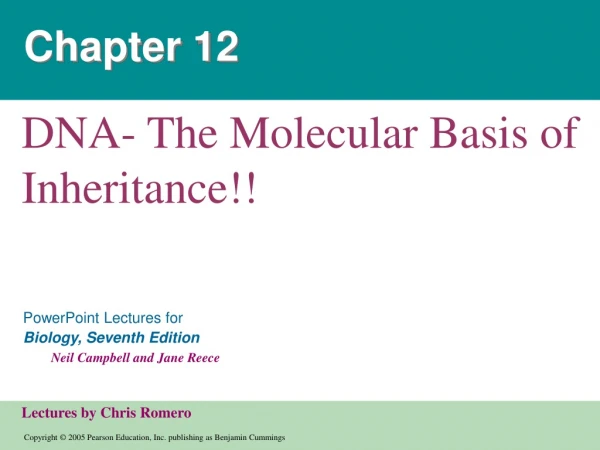
Chapter 12 DNA- The Molecular Basis of Inheritance!!
Evidence That DNA Can Transform Bacteria • The discovery of the genetic role of DNA began with research by Frederick Griffith in 1928 • Griffith worked with two strains of a bacterium, a pathogenic “S” strain and a harmless “R” strain • When he mixed heat-killed remains of the pathogenic strain with living cells of the harmless strain, some living cells became pathogenic • He called this phenomenon transformation, now defined as a change in genotype and phenotype due to assimilation of foreign DNA
LE 16-2 Mixture of heat-killed S cells and living R cells Heat-killed S cells (control) Living R cells (control) Living S cells (control) RESULTS Mouse dies Mouse healthy Mouse healthy Mouse dies Living S cells are found in blood sample
LE 16-3 Phage head Tail Tail fiber DNA 100 nm Bacterial cell
Overview: Life’s Operating Instructions • In 1953, James Watson and Francis Crick introduced an elegant double-helical model for the structure of deoxyribonucleic acid, or DNA • DNA, the substance of inheritance, is the most celebrated molecule of our time • Hereditary information is encoded in DNA and reproduced in all cells of the body • This DNA program directs the development of biochemical, anatomical, physiological, and (to some extent) behavioral traits
LE 16-5 Sugar–phosphate backbone Nitrogenous bases 5 end Thymine (T) Adenine (A) Cytosine (C) Phosphate DNA nucleotide Sugar (deoxyribose) 3 end Guanine (G)
Building a Structural Model of DNA: Scientific Inquiry • After most biologists became convinced that DNA was the genetic material, the challenge was to determine how its structure accounts for its role • Maurice Wilkins and Rosalind Franklin were using a technique called X-ray crystallography to study molecular structure • Franklin produced a picture of the DNA molecule using this technique
LE 16-6 Franklin’s X-ray diffraction photograph of DNA Rosalind Franklin
Franklin’s X-ray crystallographic images of DNA enabled Watson to deduce that DNA was helical • The X-ray images also enabled Watson to deduce the width of the helix and the spacing of the nitrogenous bases • The width suggested that the DNA molecule was made up of two strands, forming a double helix Animation: DNA Double Helix
LE 16-7 5 end Hydrogen bond 3 end 1 nm 3.4 nm 3 end 0.34 nm 5 end Key features of DNA structure Partial chemical structure Space-filling model
Watson and Crick built models of a double helix to conform to the X-rays and chemistry of DNA • Franklin had concluded that there were two antiparallel sugar-phosphate backbones, with the nitrogenous bases paired in the molecule’s interior • At first, Watson and Crick thought the bases paired like with like (A with A, and so on), but such pairings did not result in a uniform width • Instead, pairing a purine with a pyrimidine resulted in a uniform width consistent with the X-ray
LE 16-UN298 Purine + purine: too wide Pyrimidine + pyrimidine: too narrow Purine + pyrimidine: width consistent with X-ray data
Watson and Crick reasoned that the pairing was more specific, dictated by the base structures • They determined that adenine paired only with thymine, and guanine paired only with cytosine
LE 16-8 Sugar Sugar Adenine (A) Thymine (T) Sugar Sugar Guanine (G) Cytosine (C)
12-2 DNA Replication! • Since the two strands of DNA are complementary, each strand acts as a template for building a new strand in replication • In DNA replication, the parent molecule unwinds, and two new daughter strands are built based on base-pairing rules Animation: DNA Replication Overview
LE 16-9_1 The parent molecule has two complementary strands of DNA. Each base is paired by hydrogen bonding with its specific partner, A with T and G with C.
LE 16-9_2 The first step in replication is separation of the two DNA strands. The parent molecule has two complementary strands of DNA. Each base is paired by hydrogen bonding with its specific partner, A with T and G with C.
LE 16-9_3 The first step in replication is separation of the two DNA strands. Each parental strand now serves as a template that determines the order of nucleotides along a new, complementary strand. The parent molecule has two complementary strands of DNA. Each base is paired by hydrogen bonding with its specific partner, A with T and G with C.
LE 16-9_4 The first step in replication is separation of the two DNA strands. Each parental strand now serves as a template that determines the order of nucleotides along a new, complementary strand. The parent molecule has two complementary strands of DNA. Each base is paired by hydrogen bonding with its specific partner, A with T and G with C. The nucleotides are connected to form the sugar-phosphate back- bones of the new strands. Each “daughter” DNA molecule consists of one parental strand and one new strand.
Getting Started: Origins of Replication • Replication begins at special sites called origins of replication, where the two DNA strands are separated, opening up a replication “bubble” • A eukaryotic chromosome may have hundreds or even thousands of origins of replication • Replication proceeds in both directions from each origin, until the entire molecule is copied • At the end of each replication bubble is a replication fork, a Y-shaped region where new DNA strands are elongating
Elongating a New DNA Strand • Enzymes called DNA polymerases catalyze the elongation of new DNA at a replication fork • Each nucleotide that is added to a growing DNA strand is a nucleoside triphosphate • The rate of elongation is about 500 nucleotides per second in bacteria and 50 per second in human cells
LE 16-13 New strand Template strand 5¢ end 3¢ end 5¢ end 3¢ end Sugar Base Phosphate DNA polymerase 3¢ end 3¢ end Pyrophosphate Nucleoside triphosphate 5¢ end 5¢ end
Antiparallel Elongation • The antiparallel structure of the double helix (two strands oriented in opposite directions) affects replication • DNA polymerases add nucleotides only to the free 3end of a growing strand; therefore, a new DNA strand can elongate only in the 5 to 3direction
Along one template strand of DNA, called the leading strand, DNA polymerase can synthesize a complementary strand continuously, moving toward the replication fork • To elongate the other new strand, called the lagging strand, DNA polymerase must work in the direction away from the replication fork • The lagging strand is synthesized as a series of segments called Okazaki fragments, which are joined together by DNA ligase
LE 16-14 3¢ 5¢ Parental DNA Leading strand 5¢ 3¢ Okazaki fragments Lagging strand 3¢ 5¢ DNA pol III Template strand Leading strand Lagging strand Template strand DNA ligase Overall direction of replication
Priming DNA Synthesis • DNA polymerases cannot initiate synthesis of a polynucleotide; they can only add nucleotides to the 3 end • The initial nucleotide strand is a short one called an RNA or DNA primer • An enzyme called primase can start an RNA chain from scratch • Only one primer is needed to synthesize the leading strand, but for the lagging strand each Okazaki fragment must be primed separately
LE 16-15_1 Primase joins RNA nucleotides into a primer. 3¢ 5¢ 3¢ 5¢ Template strand Overall direction of replication
LE 16-15_2 Primase joins RNA nucleotides into a primer. 3¢ 5¢ 3¢ 5¢ Template strand DNA pol III adds DNA nucleotides to the primer, forming an Okazaki fragment. 3¢ 5¢ RNA primer 3¢ 5¢ Overall direction of replication
LE 16-15_3 Primase joins RNA nucleotides into a primer. 3¢ 5¢ 3¢ 5¢ Template strand DNA pol III adds DNA nucleotides to the primer, forming an Okazaki fragment. 3¢ 5¢ RNA primer 3¢ 5¢ After reaching the next RNA primer (not shown), DNA pol III falls off. Okazaki fragment 3¢ 3¢ 5¢ 5¢ Overall direction of replication
LE 16-15_4 Primase joins RNA nucleotides into a primer. 3¢ 5¢ 3¢ 5¢ Template strand DNA pol III adds DNA nucleotides to the primer, forming an Okazaki fragment. 3¢ 5¢ RNA primer 3¢ 5¢ After reaching the next RNA primer (not shown), DNA pol III falls off. Okazaki fragment 3¢ 3¢ 5¢ 5¢ After the second fragment is primed, DNA pol III adds DNA nucleotides until it reaches the first primer and falls off. 5¢ 3¢ 3¢ 5¢ Overall direction of replication
LE 16-15_5 Primase joins RNA nucleotides into a primer. 3¢ 5¢ 3¢ 5¢ Template strand DNA pol III adds DNA nucleotides to the primer, forming an Okazaki fragment. 3¢ 5¢ RNA primer 3¢ 5¢ After reaching the next RNA primer (not shown), DNA pol III falls off. Okazaki fragment 3¢ 3¢ 5¢ 5¢ After the second fragment is primed, DNA pol III adds DNA nucleotides until it reaches the first primer and falls off. 5¢ 3¢ 3¢ 5¢ DNA pol I replaces the RNA with DNA, adding to the 3¢ end of fragment 2. 5¢ 3¢ 3¢ 5¢ Overall direction of replication
LE 16-15_6 Primase joins RNA nucleotides into a primer. 3¢ 5¢ 3¢ 5¢ Template strand DNA pol III adds DNA nucleotides to the primer, forming an Okazaki fragment. 3¢ 5¢ RNA primer 3¢ 5¢ After reaching the next RNA primer (not shown), DNA pol III falls off. Okazaki fragment 3¢ 3¢ 5¢ 5¢ After the second fragment is primed, DNA pol III adds DNA nucleotides until it reaches the first primer and falls off. 5¢ 3¢ 3¢ 5¢ DNA pol I replaces the RNA with DNA, adding to the 3¢ end of fragment 2. 5¢ 3¢ 3¢ 5¢ DNA ligase forms a bond between the newest DNA and the adjacent DNA of fragment 1. The lagging strand in the region is now complete. 5¢ 3¢ 3¢ 5¢ Overall direction of replication
Other Proteins That Assist DNA Replication • Helicase untwists the double helix and separates the template DNA strands at the replication fork • Single-strand binding protein binds to and stabilizes single-stranded DNA until it can be used as a template • Topoisomerase corrects “overwinding” ahead of replication forks by breaking, swiveling, and rejoining DNA strands
Primase synthesizes an RNA primer at the 5 ends of the leading strand and the Okazaki fragments • DNA pol III continuously synthesizes the leading strand and elongates Okazaki fragments • DNA pol I removes primer from the 5 ends of the leading strand and Okazaki fragments, replacing primer with DNA and adding to adjacent 3 ends • DNA ligase joins the 3 end of the DNA that replaces the primer to the rest of the leading strand and also joins the lagging strand fragments
LE 16-16 Overall direction of replication Lagging strand Leading strand Origin of replication Leading strand Lagging strand OVERVIEW DNA pol III Leading strand DNA ligase Replication fork 5¢ DNA pol I 3¢ Primase Lagging strand Parental DNA DNA pol III Primer 3¢ 5¢
The DNA Replication Machine as a Stationary Complex • The proteins that participate in DNA replication form a large complex, a DNA replication “machine” • The DNA replication machine is probably stationary during the replication process • Recent studies support a model in which DNA polymerase molecules “reel in” parental DNA and “extrude” newly made daughter DNA molecules
Proofreading and Repairing DNA • DNA polymerases proofread newly made DNA, replacing any incorrect nucleotides • In mismatch repair of DNA, repair enzymes correct errors in base pairing • In nucleotide excision repair, enzymes cut out and replace damaged stretches of DNA
LE 16-17 A thymine dimer distorts the DNA molecule. A nuclease enzyme cuts the damaged DNA strand at two points and the damaged section is removed. Nuclease Repair synthesis by a DNA polymerase fills in the missing nucleotides. DNA polymerase DNA ligase DNA ligase seals the free end of the new DNA to the old DNA, making the strand complete.
Replicating the Ends of DNA Molecules • Limitations of DNA polymerase create problems for the linear DNA of eukaryotic chromosomes • The usual replication machinery provides no way to complete the 5 ends, so repeated rounds of replication produce shorter DNA molecules
LE 16-18 5¢ Leading strand End of parental DNA strands Lagging strand 3¢ Last fragment Previous fragment RNA primer Lagging strand 5¢ 3¢ Primer removed but cannot be replaced with DNA because no 3¢ end available for DNA polymerase Removal of primers and replacement with DNA where a 3¢ end is available 5¢ 3¢ Second round of replication 5¢ New leading strand 3¢ New leading strand 5¢ 3¢ Further rounds of replication Shorter and shorter daughter molecules
Eukaryotic chromosomal DNA molecules have at their ends nucleotide sequences called telomeres • Telomeres do not prevent the shortening of DNA molecules, but they do postpone the erosion of genes near the ends of DNA molecules
LE 16-19 1 µm
If chromosomes of germ cells became shorter in every cell cycle, essential genes would eventually be missing from the gametes they produce • An enzyme called telomerase catalyzes the lengthening of telomeres in germ cells