Download
1 / 17
260 likes | 884 Views
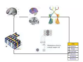
Visual Cortex. Vision Science Lectures in Ophthalmology Curtis Baker. receptive fields of retinal ganglion cells. KSJ, Fig 26-7. retina-LGN-cortex. KSJ, Fig 27-4. V1 neurons: orientation selectivity. KSJ, Fig 27-11. V1 neurons: direction selectivity. -. -. +. +. -. -. -. -. +.

E N D
Visual Cortex Vision Science Lectures in Ophthalmology Curtis Baker
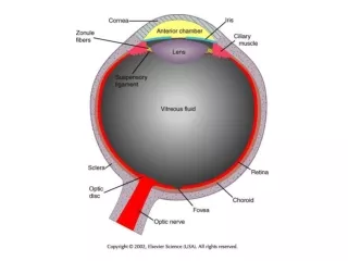
receptive fields of retinal ganglion cells KSJ, Fig 26-7
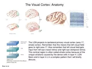
retina-LGN-cortex KSJ, Fig 27-4
V1 neurons: orientation selectivity KSJ, Fig 27-11
- - + + - - - - + + - - - - + + + + - - - - - - - - + + - - + + - - - - - - + + - - - - + + - - - + - + + - + - - - - - - - + + - - + - + - - - neurons as stimulus filters wrong spatial frequency simple cell receptive field wrong orientation optimal grating
ocular dominance columns KSJ, Fig 27-16
CCD Camera Population responses: optical imaging Stimulus Generator Illuminator Real-time Video Processor Data Collection and Analysis (courtesy of Chang’an Zhan)
orientation columns KSJ, Fig 27-14
cytochrome oxidase blobs KSJ, Fig 27-15 • input from LGN koniocellular layers • in centers of ocular dominance columns • receptive fields: non-oriented, colour-opponent
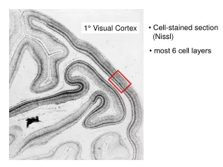
retinotopic map in V1 KSJ, Fig 27-9
retinotopic maps from fMRI Sereno et al, 1995
field sign maps Sereno et al, 1995
extrastriate visual areas larger receptive fields, less retinotopy functional specializations single units: Zeki, V4 vs MT/V5 brain imaging: stimuli that selectively activate (e.g., motion/flicker -> MT/V5) KSJ, Fig 25-9
dorsal & ventral streams: many extrastriate areas KSJ, Fig 28-2
dorsal and ventral streams: neurophysiology KSJ, Fig 25-12
effects of damage striate cortex (V1) -> blindness V2 -> "quadrant" blindness other extrastrate areas -> selective losses, e.g.: MT / V5 -> motion-blindness FFA (fusiform face area) -> prosopagnosia fusiform cortex ("colour area" ?) -> achromatopsia