Download
1 / 18
180 likes | 387 Views
Receptive Field Dynamics in Adult Primary Visual Cortex. Group A3 Presenters: Anastasia Christopher, Carol Rego , Sarah McNeil Technical Experts: Bonnie Chan, Herman Gill, Marisa Leung. Anastasia Christopher. Table of Contents. Brief overview of Module 3

E N D
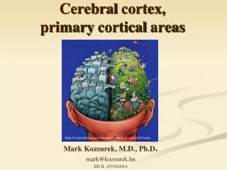
Receptive Field Dynamics in Adult Primary Visual Cortex Group A3 Presenters: Anastasia Christopher, Carol Rego, Sarah McNeil Technical Experts: Bonnie Chan, Herman Gill, Marisa Leung
Anastasia Christopher Table of Contents • Brief overview of Module 3 • Background information and important definitions • Anatomy of the brain • Lateral Geniculate Nucleus • Method • Results • Primary Visual Cortex • Method • Results • Conclusion
Anastasia Christopher Receptive Field Dynamics in Adult Primary Visual Cortex By: Charles Gilbert and Torsten Wiesel
Anastasia Christopher Important Definitions • Cortical Topography • Binocular Retinal Lesion • Scotoma Receptive Field Size
Anastasia Christopher Is the locus of change in regards to sensory input located at the cortical level (Primary Visual Cortex) or at a prior stage in the sensory pathway (Lateral Geniculate Nucleus)?
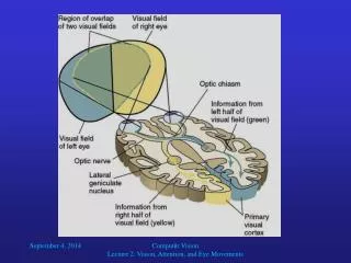
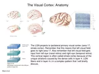
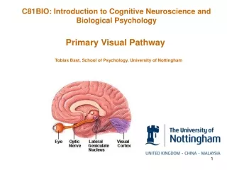
Anastasia Christopher Lateral Geniculate Nucleus (LGN)
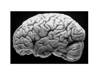
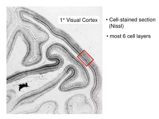
Anastasia Christopher Primary Visual Cortex
Carol Rego LGN - Methods • Studied 2 cats and 1 monkey • Topographical mapping of the LGN • Multiple electrode penetrations across the LGN • Two injections of retrograde tracers in V1
Carol Rego LGN – Electrode Readings • Large silent area about 1mm in diameter • Shift in RF position
Sarah McNeil Primary Visual Cortex - Methods • Studied 4 cats and 6 monkeys • RF maps were made using vertical electrode penetration • RF maps were made at the same sites before, immediately after, and 2 months after making the lesion
Sarah McNeil Primary Visual Cortex - Results • Immediately after: • Inactivity in original sites • 5X larger sites • Centrifugal shift Before lesion Immediately after lesion
Sarah McNeil Primary Visual Cortex - Results • 2 months after: • RF field size shrunk • 5˚ centrifugal shift in position • Area of activity expanded beyond affected area Before Lesion 2 Months After Lesion
Sarah McNeil • Initial shock • Long term consolidation
Anastasia Christopher Is the locus of change in regards to sensory input located at the cortical level (Primary Visual Cortex) or at a prior stage in the sensory pathway (Lateral Geniculate Nucleus)?
Anastasia Christopher Conclusion • Brain is high in plasticity • Compensation of damaged tissue as a result of binocular retinal lesions does not take place in LGN • Takes place at the cortical level of V1