Download
1 / 34
340 likes | 508 Views
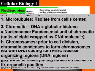
Cellular Biology. School of Life Sciences Shaanxi Normal University. 1. CHAPTER 6 MITOCHONDRION AND CHLOROPLAST. I. Mitochondrion Structure and distribution:

E N D
Cellular Biology School of Life Sciences Shaanxi Normal University 1
CHAPTER 6 MITOCHONDRION AND CHLOROPLAST
I. Mitochondrion Structure and distribution: Usually, mitochondrion is like a particle or rod and others composed of proteins (65-70%) and lipids (25-30%) at 0.5~1μm diameter and 1.5~3.0μm length. The mitochondria in the cells of pancreas can be 10~20μm at length called huge mitochondrion. The number of mitochondrion in a cell can be hundreds to thousands, and less in plant cell than in animal cell because of chloroplast. Some unicellular organism contains 500,000 mitochondria inside, but mammalian erythrocytes contain no any mitochondrion inside. Mitochondrion can migrate in cell along micro tube, and motorprotein supplies energy for that.
1. Outer membrane Outer membrane contains lipids (40%) and proteins (60%). There are the hydrophilic tunnels composed of porin that allows the molecules lighter than 5KD passed through. 2. Inner membrane Inner membrane contains more than 100 types of polypeptide with a low permeability. The electron transmission chain of oxidative phosphorylation is located in inner membrane. Cytochrome C reductase is the marker enzyme for inner membrane. Inner membrane can be pleated into inside to form cristae. The cristaes enlarge the area of inner membrane to 5 – 10 folds. Cristae can be two types of shape: lamella or tube. Elementary particles are located on cristae, and composed of head part (F1 conjugate factor) and elemantary part (F0 conjugate factor). F0 inserts into inner membrane. 3. Intermembrane space It is between inner membrane and outer membrane with 6-8nm width. Adenylate kinase is the marker enzyme for intermembrane space.
Inner membrane Photo of mitochondrion Outer membrane Intermembrane space Lamella critae
Matrix A model structure of mitochondrion
4. Matrix The area surrounded by cristae, intermembrane space and inner membrane. Excepting glycolysis (in plasma), other bio-oxidation reactions are carried out in mitochondrion (in matrix). Malic dehydrogenase (MDH) is the marker enzyme for the matrix of mitochondrion. The matrix contains a complete system for transcription and translation including mitochondrion DNA (mtDNA), 70s ribosome, tRNA, rRNA, and DNA polymerase. Tube cristae Matrix
The molecule basis of oxidative phosphorylation Electronic vectors for the respiratory chain: 1. Nicotinamide adenine dinucleotide (NAD): NAD is a coenzyme for many dehydrogenases and linked to tricarboxylic acids cycle. NAD present H+ to flavoprotein. 2. Flavoprotein. 3. Cytochromes: Types: a、a3、b、c、c1. 4. Three Copper atoms on the protein located on inner membrane. 5. Ferredoxin. 6. Coenzyme Q. A model structure for ferredoxin
ATP synthetase: Molecule weight: 500KD. Two parts: a spherical head (F1), and the basement (F0) inserted membrane. Each liver cell mitochondrion contains 15,000 molecules of ATP synthetase. Each ATP synthetase can make 100 ATP molecules per second. For the detail about ATP synthetase, see your biochemistry text book.
The inhibitors for oxidative phosphorylation: 1.Inhibitors for electronic transmission ① Inhibit NADH→CoQ: amytal, rotenone, and piericidin. ② Inhibit Cyt b→Cyt c: actinomycin A. ③ Inhibit cytochrome oxidase→O2: CO, CN, NaN3, and H2S. 2.Inhibitors for phosphorylation Oligomycin and dicyclohexyl carbodiimide (DCC) can bind to F0 to block the H+ tunnel and inhibit synthesis of ATP. 3.Uncoupler Some reagents or medicines can separate the oxidation and phosphorylation, oxidation is continued but phosphorylation stopped. The uncoupler can cause body temperature increased. Uncouplers include DNP, FCCP, thermogenin, and aspirin.
Mitochondrion is semiautonomous Mitochondrion contains its whole system for DNA replication, transcription, and translation. The system includes mtDNA, RNA, DNA polymerase, RNA polymerase, tRNA, ribosome, and others. That means mitochondrion has the genetic way for itself. So, we say that mitochondrion is a semiautonomous organelle. mtDNA is double strands and circle molecule as the below: Heavy chain (H) Light chain (L) The genes on mtDNA are located very closely without introns. Each mitochondrion contains several mtDNA molecules, and the length for each is about 16-20kb in animal. Most of gene are transcripted from H chain. The genes on H chain encode two rRNAs, 14 tRNAs, and 12 polypeptides. The genes on L Chain encode other 8 tRNAs and 1 peptide. The genes on mtDNA linked together, or overlapped. Almost each reading frame has no any region that is not translated. Many of them have no stop codon, and end as T or TA. The stop codon will be added during the modification after transcription.
Mitochondrion was originated from a bacterium that parasited in cell probably because mitochondrion has many features are almost same to bacteria, for examples, morphology, staining, chemical components, genetic system, and others. The following genetic features are same to a bacterium’s: ① circle DNA without intron. ② 70S ribosome. ③ RNA polymerase can be inhibited by EB, not by actinomycin D. ④ Sensitive to chloromycetin that inhibits bacterial protein synthesis, not to actidione that inhibits the cellular protein synthesis. The genetic codons of mammalian mtDNA are different from universal genetic codons: ① UGA is not a stop codon here, it is a codon for Tryptophan. ② Methionine is encoded by codon AUG, AUA, AUU and AUC. ③ AGA and AGG are not codons for arginine, they are stop codons here. There are 4 stop codons in mitochondrion: UAA, UAG, AGA, and AGG. mtDNA is transferred to new generation from parental generation with a matrilinear inheritance way, and its mutation rate is higher than nucleus DNA (nDNA) without efficient repairing function. So, mtDNA is easy to be mutated and cause mutation genetic diseases, such as Leber optic nerve disease (optic nerve denaturation and atrophy) and myoclonus epilepsy (convulsive seizure and loss of consciousness).
Proliferation of mitochondrion Mitochondrion is proliferated by the cleavage styles include: 1. Septate division: The mitochondrion membrane forms a ligature ditch rounding the middle of membrane, then separated to two new mitochondria.
2. Contracting division: The ligature ditch become slender and is pulled to longer further, the divided off to new mitochondria. 3. Budding division: Germinated firstly, the small mitochondrion will fall off from its mother mitochondrion, grow up and develop to a new mitochondrion. Contracting division of mitochondrion
II. Chloroplast We can say that all energy utilized by every life event is originally from sun. But, how to utilize and transform it? We, human body can not take energy from sun, but we have to use it by other way. Chloroplast plays key role here! Chloroplast take and transform the sun energy for plant growth, animals take energy and nutrition from plants (or other animal). As a energy transformer, chloroplast can combine carbon dioxide and water to form sugar and release oxygen. So, the photosynthesis of green plants is the basic energy resource for all bio organs including human being in the world. Sunlight 6CO2+6H2O C6H12O6+6O2 Chloroplast Structure: The size of chloroplast is about 5~10um × 2~4um × 2~3um. 50 – 200 chloroplasts are contained in each plant cell usually. The size and shape are different in different plant species. Chloroplast is composed of envelope, thylakoid and stroma. Chloroplast contains 3 types of membranes (outer membrane, inner membrane and thylakoid membrane) and 3 types of cavities (intermembrane space, stroma cavity and thylakoid space).
Envelope: Envelope is composed of bilayer of lipid membrane with a 10~20nm intermembrane space. The outer layer allows almost all large or small molecules pass through, but the inner layer allows molecules pass through selectively. Thylakoid: Thylakoid is a vesicle formed by a monolayer membrane with some components of photosynthetic pigments and electronic transmission, we call this layer as photosynthetic membrane. 10 – 100 thylakoids are overlapped together to form a basic particles, so we call these thylakoids as basic particle thylakoids. Each chloroplast contains 40 – 60 basic particles. The thylakoids that located between basic particles and did not overlap together are called stroma thylakoids that form stroma lamella. The basic particles are linked by stroma thylakoids. So, all thylakoids form a closed system. The membrane of thylakoid is composed of protein and lipid(60:40). The light energy is transformed into chemical energy on thylakoid membrane. The proteins contained in the thylakoid membrane are the complex of cytochromeb6/f, flavoprotein, complex of photosystem I, complex of photosystem II, and others.
Stroma: Stroma is the area between inner membrane and thylakoid. The components of stroma include: Enzymes associated with carbon assimilation, The system for protein synthesis [ chloroplast DNA (ctDNA), RNAs, ribosome], and others. The mechanism of photosynthesis: Photosynthesis is a course for the transformation of energy and substance: Electronic transmission Light energy Electric Chemical energy Store in (Sun energy) power (ATP and NADPH) sugars The course can be divided as two parts: light reaction (the reaction needs light) and dark reaction (the reaction does not need light). The photosynthetic pigments and electronic transmission components: Photosynthetic pigments: Thylakoid contains two pigments: green chlorophyll and red carotinoid (3:1). Chlorophyll includes two types: chlorophyll a and b. All chlorophylls and carotinoid are embedded in thylakoid membrane. So, thylakoid membrane is a very important place where the photosynthesis and energy exchange are carried out.
Light harvesting complex Light harvesting complex is composed of about 200 chlorophylls and some peptides. Most of chlorophyll a and all chlorophyll b can capture light, they are called as antenna pigment. Carotinoid and xanthophyll are the helper pigments. Antenna pigments transfer the light energy to central chlorophyll by a resonance energy transmission way.
Actually, there are 3 plant pigments in thylakoid: green chlorophyll, red carotene plus carotinoid, and yellow xanthophyll. The 3 pigments form 3 basic colors to figure out so colorful and beautiful world to us that we can not love her enough forever! Photosystem II (PSⅡ): PS II is called as P680 also. PS II includes 12 peptide chains at least. PS II is located in thylakoid membrane, and contains one light-hawesting comnplex Ⅱ (LHC Ⅱ), one central chlorophyll, and one oxygen evolving complex. D1 and D2 are the key peptide chains combined to P680、pheophytin and plastoquinone. Cytochrome b6/f complex (cyt b6/f complex): Cytochrome b6/f complex exists in dimer style. Each monomer contains four subunits: cytochrome b6 (b563), cytochrome f, ferredoxin, and subunit Ⅳ. Photosystem I (PS I): PS I is called as P700 also. PS I is located in stroma and thylakoid membrane. PS I is composed of light harvesting complex I and a reaction center formed by some special chlorophylls, electron vectors.
Light capturing reaction and electron transmission: Light capture: Electron trnasfer: Light energy P680 (ground state) P680e+(excited state) Pheophytin QA on D2 QB on D1 Plastoquinone of PS II Complex of cytochrome b6/f Cu2+ on plastocyanin P700 of PS I A0 A1 4Fe-4S Ferredoxin NADP+ NADPH The “Z” way of electron transmission above is called as non-cycle photosynthetic phosphorylation. Both ATP and NADPH are released out here.
If the NADP+ is inefficient in plant, electron will cycle in PS I to form ATP only, no NADPH synthesized at this time. We call this transmission as cycle photosynthetic phosphorylation shown as the fig below. Cycle photosynthetic phosphorylation
The cooperation of PS I and PS II (The fig just show you the “Z” way)
Photosynthetic phosphorylation: Just like the described as above, a pair of electrons is transferred from P680 to NADP+, this transmission will put 4 H+ ions into the cavity of thylakoid making the pH here low (about 5) and forming H+ force. Under the ATP synthetase enhancing, the H+ force will push ADP combined by Pi to form ATP. Oxidative phosphorylation and photosynthetic phosphorylation
Semiautonomy of chloroplast Like mitochondrion, chloroplast is the energy station in plant cell. Chloroplast is a semiautonomous organelle also. Chloroplast DNA (ctDNA) is cycle DNA with 200Kb-2500Kb length usually. Like mitochondrion, chloroplast synthesizes the some of proteins that chloroplast needs. Other needed proteins must be synthesized in cell plasma. So, we say that chloroplast is a semiautonomous organelle also. The proliferation of chloroplast The proliferation of chloroplast is very similar to the septate division of mitochondrion. The chloroplast membrane forms a ligature ditch rounding the middle of membrane, then separated two new chloroplasts. Chloroplast cleavage is easier to be found in baby plant or leaf than in adult plant or leaf usually. A adult chloroplast will not start division again almost.
Directive transportation of proteins in mitochondrion and chloroplast Transportation of mitochondrion proteins Most of mitochondrion proteins are synthesized in plasma and transported into mitochondrion directionally. The signal sequence (leader sequence, presequence or transit-peptide) at N terminal of protein will lead the transportation specifically. After the transportation, the signal sequence will be cut off by signal peptidase. We call this splicing as posttranslation modification.
The protein transportation signal sequence is not specific to target protein. Any protein with the signal sequence can be transported into mitochondrion. Many protein complexes are associated with the protein transportation: These complexes are the translocators actually including the follows:: ①TOM complex: Protein can enter intermembrane space by this complex. The important human TOMs are TOM34, TOM40, TOM22, TOM7, TOM6, and Tom5. The tunnel of TOM complex is called as general import pore (GIP). ②TIM complex: TIM23 can transport protein to stroma or insert some proteins into inner membrane. TIM22 can insert the transportation proteins for metabolism needed substances into inner membrane. ③OXA complex: OXA can insert both the proteins synthesized by mitochondrion and the proteins passed through TOM/TIM into inner membrane. The proteins entered outer membrane contain a N terminal signal that will not be cut off and will be inserted into outer membrane by TOM complex. For stroma proteins, they have to be transported into intermembrane space by TOM complex firstly, then inserted into stroma by TIM complex. As another choice, they can enter stroma by the cooperation of TOM and TIM at the site where the mitochondrion inner and outer membranes are touched each other.
The complex of TOM and TIM (Proteins can pass through outer membrane and inner membrane here with one step only)
The touched sites of the outer membrane and inner membrane of mitochondrion
Protein enter inner membrane and intermembrane space: A. The protein contains two signal sequences enter stroma by first signal for TOM/TIM23, then, inserted into inner membrane by second signal for OXA complex. B. The proteins entered intermembrane space can be inserted inner membrane by the stop-transfer sequence for TIM23. C. The inserted proteins can be modified by the protease as soluble proteins. D. The transporter for metabolized substance can be inserted into inner membrane byTIM22.