Download
1 / 1
10 likes | 103 Views
PROTEOMICS OF REDIVAC FLUID: COMPARATIVE ANALYSIS OF PROTEIN CLUSTERS IN PERITONEAL LAVAGE FLUID AND GYNAECOLOGICAL CANCER

E N D
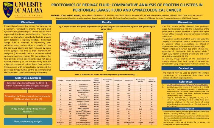
PROTEOMICS OF REDIVAC FLUID: COMPARATIVE ANALYSIS OF PROTEIN CLUSTERS IN PERITONEAL LAVAGE FLUID AND GYNAECOLOGICAL CANCER EUGENE LEONG WENG KONG1, RAMARAO SERIRAMALU2, PUTERI SHAFINAZ ABDUL RAHMAN2,3, NOOR AZMI MOHAMAD ADENAN1 AND ONN HAJI HASHIM2,3.1Department of Obstetrics and Gynaecology, 2Department of Molecular Medicine, Faculty of Medicine, 3University of Malaya Centre for Proteomics Research, 50603 Kuala Lumpur. Objectives Results Discussions Gynaecological cancer is a cancer that develops in a woman’s reproductive organs. The signs and symptoms for gynaecological cancer remain to be vague and thus hinder early detection. Therefore search for biomarkers using body fluids to provide early detection is urgently needed. Peritoneal lavage fluid is obtained at laparotomy pre-definitive surgery when saline is introduced into the peritoneal cavity, and then removed by clean sterile syringe. The fluid is then examined for malignant (cancer) cells under a procedure called peritoneal washing cytology [1]. Interestingly, the fluid and its protein constituents have not been studied previously. In the present study, we have subjected proteins of redivac fluids from patients (with consent) with gynaecological cancer and the peritoneal lavage fluid as control to 2-dimensional electrophoresis (2-DE) and mass spectrometry. • The 2DE protein profile obtained from peritoneal lavage fluid is comparable with the redivac fluid of the gynaecological patient. However, a significantly higher number of low molecular proteins were resolved in the redivac fluid. • The proteins identified in Table 1 mainly falls under the category of acute phase proteins. Acute phase proteins are proteins whose concentrations are altered in response to trauma, infection and inflammation[3]. • Visual comparison between 2DE profile shows over-expression of haptoglobin (11), Ig alpha-1 chain C region (15), vitamin D-binding protein (16) and serum albumin fragments (21, 22, 23). • At present, image analysis of the expression of proteins clusters from both group of samples are performed by using Image Master Platinum version 7.0. Fig 1. Representative 2-DE profile ofperitoneal lavage fluid (left) and redivac fluid from a patient with gynaecological cancer (right). Conclusions The method may be used to analyse the protein compositions of post-operative drain fluids from various cohorts of surgical patients. Table 1. Maldi ToF/ToF results obtained for proteins spots detected in Fig. 1. Materials & Methods References Collection of peritoneal lavage fluid (n=3) & redivac fluid from patients with gynaecological cancer (n = 3) http://hepatitis.about.com/od/pqr/g/PeritonealFluid.htm Abdul-Rahman, P. S., Lim, B. K., & Hashim, O. H. (2007). Expression of high-abundance proteins in sera of patients with endometrial and cervical cancers: Analysis using 2-DE with silver staining and lectin detection methods. Electrophoresis, 28(12), 1989-1996. Gruys, E., Toussaint, M. J., Upragarin, N., Van, E. A., Adewuyi, A. A., Candiani, D., et al. (2005). Acute phase reactants, challenge in the near future of animal production and veterinary medicine. J Zhejiang Univ Sci B, 6(10), 941-947. Separation by 2-dimensional electrophoresis (2-DE) and silver staining [2] Image analysis using Image Master Platinum 7 Acknowledgement • This work was funded by research grant UMRG RG278/10HTM • from the University of Malaya, Kuala Lumpur. • Mass spectrometry analysis was performed with the access to • the Proteomic facility at the Medical Biotechnology Laboratory, • Faculty of Medicine, UM. • Special thanks to Ms. Nazirah Abrahim for her assistance in sample collection and poster design. Mass spectrometry analysis