Download
1 / 43
430 likes | 542 Views
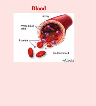
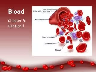
Blood. Blood accounts for approximately 8% of body weight The pH of blood is 7.35–7.45 Temperature is 38 C, slightly higher than “ normal ” body temperature Average volume of blood is 5–6 L for males, and 4–5 L for females RBC circulate for approximately 4 months.

E N D
Blood • Blood accounts for approximately 8% of body weight • The pH of blood is 7.35–7.45 • Temperature is 38C, slightly higher than “normal” body temperature • Average volume of blood is 5–6 L for males, and 4–5 L for females • RBC circulate for approximately 4 months.
Functions of Circulatory System • Transport • O2, CO2, nutrients, wastes, hormones, and heat • Protection • WBCs, antibodies, and platelets • Regulation fluid volume in the circulatory system • When the blood has a high osmolarity water will leave the tissues and go into blood. • This is why excessive consumption of certain electrolytes raises BP • low osmolarity • When blood has a low osmolarity excessive fluid follows an osmotic gradient into the tissues which may result in edema
Erythrocytes (RBCs) • Biconcave discs, anucleate, essentially no organelles • Filled with hemoglobin (Hb) • a protein that functions in gas transport • Contain the plasma membrane protein spectrin and other proteins that: • Give erythrocytes their flexibility • Allow them to change shape as necessary
Production of Erythrocytes • Hematopoiesis – blood cell formation • Hematopoiesis occurs in the red bone marrow of the: • Axial skeleton and girdles • Epiphyses of the humerus and femur • Hemocytoblasts give rise to all formed elements
Hormonal Control of Erythropoiesis • Chemoreceptors in the kidneys detect a decrease in P02 • They respond by secreting the hormone Erythropoietin. (EPO) • (EPO) is triggered by: • Hypoxia( low oxygen levels) due to decreased RBCs • anemia • Decreased oxygen availability • High altitude, pulmonary infections • Increased tissue demand for oxygen • exercise • Erythropoesis increases the: • RBC count in circulating blood • Oxygen carrying ability of the blood
Erythropoietin Mechanism Imbalance Start Normal blood oxygen levels Stimulus: Hypoxia due to decreased RBC count, decreased availability of O2 to blood, or increased tissue demands for O2 Imbalance Increases O2-carrying ability of blood Reduces O2 levels in blood Erythropoietin stimulates red bone marrow Kidney (and liver to a smaller extent) releases erythropoietin Enhanced erythropoiesis increases RBC count
Erythrocyte Disorders • Polycythemia - an excess of RBCs • primary polycythemia (Polycythemia Vera ) • cancers of erythropoietic cell line in red bone marrow • secondary polycythemia (ENVIORMENTAL) • from dehydration, emphysema, high altitude, or physical conditioning and blood doping • Dangers of polycythemia • increased RBC’s will result in increases in: • blood volume (water following their osmotic gradient) • Viscosity (stickiness ,thicker) heart works harder greater resistance • Blood pressure (Both increases in vessel resistance and volume will increase the pressure in the vessel. • can lead to embolism, stroke or heart failure
Diagnostic Value In Examining RBCs • Diabetes: Chronically elevated BGLs will increase glycosylated hemoglobin ( A1c). • This reflects average BGL over the several months. • Essential fatty acid composition: Looks at composition of fatty acids that make up cell membranes. • Low omega 3 and high omega 6 and saturated fatty acids predispose you to inflammation and disease. • Magnesium levels: measures mag levels inside the RBCs opposed to just looking at serum levels. • Signs and symptoms of magnesium deficiency include fatigue, weakness, heart disturbances, mental confusion, muscle cramps, loss of appetite, and insomnia.
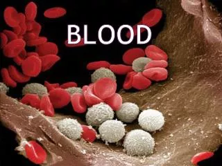
Leukocytes (WBCs) • Leukocytes, the only blood cells that are considered complete cells: • Are less numerous than RBCs and only make up 1% of the total blood volume • 1/750 ratio to RBC • ( Buffy white coat) • Can leave capillaries via diapedesis and move through tissue spaces in response to bacterial or viral invasion
Leukopoiesis Fig. 18.18
Platelets • Small fragments of megakaryocyte cytoplasm • Normal Count - 130,000 to 400,000 platelets/L • Functions • secrete clotting factors and growth factors for vessel repair • initiate formation of clot-dissolving enzyme • phagocytize bacteria • chemically attract neutrophils and monocytes to sites of inflammation
Genesis of Platelets • The stem cell for platelets is the hemocytoblast • The sequential developmental pathway from hemocytoblast to megakaryocytes. • These cells produce platelets by • Pieces of cytoplasm splits off (cell fragments) that enter bloodstream as platelets (live for 10 days) • some stored in spleen
Hemostasis • Normally blood will flow smoothly though an intact artery. If there is a disruption in the endothelium; hemorrhaging may result. The artery will protect itself through a process called hemostasis. • It involves 3 major phases
Hemostasis - Vascular Spasm The initial response of the damaged blood vessel is to constrict preventing additional blood lose. This is accomplished through activation of: pain receptors : directly innervate constrictors. smooth muscle injury results in reflexive spasm platelets release serotonin (vasoconstrictor)
Hemostasis -Platelet Plug Formation Platelet plug formation: broken vessel exposes collagen which attracts platelets Platelets form pseudopods enabling them stick to damaged vessel. Platelets produce releasing a variety of substances serotonin is a vasoconstrictor ADP attracts more platelets (positive feedback)
Hemostasis - Coagulation • Clotting - most effective defense against bleeding • Procoagulants (clotting factors) are present in plasma undergo series of biochemical reactions. • activate one factor and it will activate the next to form a reaction cascade. • Extrinsic pathway : initiated by damaged tissues • Intrinsic pathway: platelet plug produces clotting factors. • Both pathways lead to the production of factor X • The final result is conversion of fibrinogen into fibrin
Coagulation Pathways • Extrinsic pathway • initiated by tissue thromboplastin • Intrinsic pathway • initiated by factor XII • Both produce factor X which leads to: • Prothrombin activator: converts of fibrinogen to Fibrin • Clotting requires many steps to ensure clots don’t happen without cause.
Hemostasis Disorders: Bleeding Disorders • Thrombocytopenia – condition where the number of circulating platelets is deficient. • Patients show petechiae (small purple blotches on the skin) due to spontaneous, widespread hemorrhage. • Caused by suppression or destruction of bone marrow (e.g., malignancy, radiation) • Platelet counts less than 50,000/mm3 is diagnostic for this condition. • Treated with whole blood transfusions. • Hemophilia • Genetic lack of any clotting factor affects coagulation
Coagulation Disorders • What effect does stress have on platelet aggregation? • Epinephrine increases likely of forming clots by making the platelets more sticky. • Thrombosis - abnormal clotting in unbroken vessel which will narrow the lumen for blood to flow . • most likely to occur in leg veins of inactive people because stagnant blood tends to form clots. • The formation of a dangerous thrombi can block circulation resulting in tissue death • Coronary thrombosis – thrombus in blood vessel of the heart • Embolism – The thrombus over time can break of and travel throughout the in a vessel until it gets stuck. • Infarction may occur if clot blocks blood supply to an organ (MI or stroke) • pulmonary embolism - clot may break free, travel from veins to lungs • Cerebral emboli results in CVA (cerebral vascular accidents) stroke.
Lymphatic System • Immunity • fluids from all capillary beds are filtered • immune cells stand ready to respond to foreign cells or chemicals encountered • Lipid absorption • Lacteals in small intestine absorb dietary lipids • Fluid recovery • absorbs plasma proteins and fluid (2 to 4 L/day) from tissues and returns it to the bloodstream • interference with lymphatic drainage leads to severe edema
Immunity: Two Intrinsic Defense Systems • The function of the immune system is to protect the body from pathogens. • It is composed of 2 major divisions • Innate (nonspecific) • The body’s first line of defense system consists of: • intact skin and mucosa 2. The inflammation process 3. Macrophages such as monocytes which phagocytose foreign invaders. 4. NK cells. The bodies nonspecific assassins • Acquired (specific) system • Following ingestion of a pathogen by a macrophage lymphocytes become sensitive to that specific pathogens • B-lymphocytes function in the body fluid such as blood • T lymphocytes work in infected cells. • takes longer to react than the innate system • this system has memory
Inflammation: Tissue Response to Injury • The inflammatory response is triggered whenever body tissues are injured • Prevents the spread of damaging agents to nearby tissues • Disposes of cell debris and pathogens • Sets the stage for repair processes • The four cardinal signs of acute inflammation are redness, heat, swelling, and pain
Inflammatory Response: Phagocytic Mobilization Positivechemotaxis 4 Inflammatory chemicals diffusing from the inflamed site act as chemotactic agents Neutrophils enter blood from bone marrow 1 Diapedesis 3 Margination 2 Endothelium Basal lamina Capillary wall
Natural Killer (NK) Cells • They are considered part of the non specific immune system • Natural killer cells: • Are a small, distinct group of large granular lymphocytes • React nonspecifically and eliminate cancerous and virus-infected cells • Kill their target cells by releasing perforins and other cytolytic (cell- lysing) chemicals • Secrete potent chemicals that enhance the inflammatory response
Adaptive Immune Defenses • The adaptive immune system is antigen-specific, systemic and has memory. The adaptive (specific) immune system: • Recognizes specific foreign substances (Antigens) : • Substances that can mobilize the immune system and provoke an immune response • This the body sees as foreign (bad) • Acts to immobilize, neutralize, or destroy these antigens • Amplifies inflammatory response and activates complement • It has two separate but overlapping arms of defense. • Humoral, or antibody-mediated immunity • B lymphocytes – oversee humoral immunity • Extracellular (antigens)pathogens bacteria • Cellular, or cell-mediated immunity • T lymphocytes – non-antibody-producing cells that constitute the cell-mediated arm of immunity • intracellular antigens such viruses and cancer
Lymphocytes • Immature lymphocytes released from bone marrow are essentially identical • Whether a lymphocyte matures into a B cell or a T cell depends on where in the body it becomes Immunocompetent (Functional) • B cells mature in the bone marrow • Humoral division • T cells mature in the thymus • Cellular division
Cell-Mediated Immune Response • Since antibodies are useless against intracellular antigens, cell-mediated immunity is needed • Two major populations of T cells mediate cellular immunity • CD4 cells (T4 cells) are primarily helper T cells (TH) • CD8 cells (T8 cells) are cytotoxic T cells (TC) that destroy cells harboring foreign antigens • Other types of T cells are: • Suppressor T cells (TS) Turn down immune response once threat is over. • Memory T cells : reduce response time of future attacks.
Antigen Presenting Cell • Once a macrophage has eaten an antigen it becomes an Antigen-presenting cells (APCs): • Do not respond to specific antigens • They present antigen to TH-cell which alerts both B and T cells of the specific antigen.
Helper T Cells (TH) • Regulatory cells that play a central role in the immune response • Once primed by APC presentation of antigen, they: • Chemically or directly stimulate proliferation of other T cells • Stimulate B cells that have already become bound to antigen • Without TH, there is no immune response • AIDS cripples the immune system by effecting the # of TH cells.
Role of Helper T Cells • Secretes interleukins (chemical messages between cells to mobilize the response!) • attract neutrophils, NK cells, macrophages • stimulate phagocytosis • stimulate T and B cell mitosis and maturation • Coordinate humoral and cellular immunity
Cytotoxic T Cell (Tc) • TC cells, or killer T cells, are the only T cells that can directly attack and kill other cells • They circulate throughout the body in search of body cells that display the antigen to which they have been sensitized • Their targets include: • Virus-infected cells • Cells with intracellular bacteria or parasites • Cancer cells • Foreign cells from blood transfusions or transplants
TC cells: Bind to the target cell and release perforins into its membrane perforin causes cell lysis Mechanisms of Tc Action
Humoral Response • The B-cells produce soluble antibodies that interact in extracellular environments such as: • body secretions • tissue fluid • blood • lymph • Antibodies surround and incapacitate the antigens by • Preventing viruses from entering the cell • Neutralize release of endotoxins from bacteria • Surrounds antigens allowing phagocyte time to engulf them.
Humoral Immunity • B-cells are presented with an antigen directly or by T-helper cell • This activated clone B-cells with a specific antigen receptor to identify the specific antigen • The clones produce plasma cells which will produce the antibodies specific for that antigen. • Antigen will become surrounded by antibodies disabling it allowing for a phagocyte to come and finish it off. • Some B-Cells will become memory B-Cells which circulate until the next time the body is presented with that antigen. • This is why the body responds faster the next time you are exposed
Blood And Immunity Screen • Skin color changes • Shortness of breath • Blood pressure changes • Dizziness • Body weight changes • Easy bruising, bleeding • Sensory changes in hands and feet • Swollen lymph nodes • Fever • Changes in heart rate • Autonomic dysfunction • Anemia (sickle cell ) • Leukemia hx of blood transfusions.