Download
1 / 103
1.03k likes | 1.05k Views
Diseases of the Respiratory System. Prepared by Dr. Panchajani.R. Overview. Organs of the Respiratory System Bronchial tubes Larynx Lungs Nose Pharynx Trachea. Respiratory System Consists of 6 Major Organs. Nose Pharynx Larynx Trachea

E N D
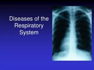
Diseases of the Respiratory System Prepared by Dr. Panchajani.R
Overview • Organs of the Respiratory System • Bronchial tubes • Larynx • Lungs • Nose • Pharynx • Trachea
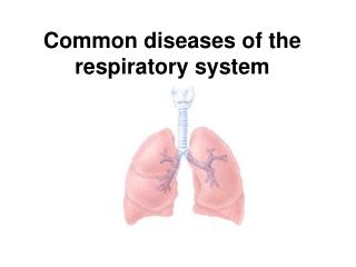
Respiratory System Consists of 6 Major Organs • Nose • Pharynx • Larynx • Trachea • Bronchial tubes • Lungs All function together to perform respiration
Respiratory System • Body cells require constant exchange of fresh oxygen and removal of carbon dioxide. • Respiratory system works in conjunction with cardiovascular system. • Process must be continuous.
3 Distinct Parts of Respiration • Ventilation – flow of air between outside and lungs • Inhalation – flow of air into lungs; brings fresh oxygen • Exhalation – flow of air out of lungs; removes carbon dioxide
External Respiration • Exchange of oxygen and carbon dioxide in lungs • Gases diffuse in opposite directions • Between air sacs of lungs and bloodstream • Oxygen enters bloodstream • Carbon dioxide leaves bloodstream.
Internal Respiration • Oxygen and carbon dioxide exchange at cellular level • Delivered to tissues • Necessary for metabolism • Referred to as tissue breathing
Exchange of gases between lungs and blood. High concentration of CO2 in blood capillary to alveolus diffuses into alveolus. High concentration of O2 in alveolus diffuses into blood capillary leaving lung.
Nose and Nasal Cavity • Beginning of ventilation process • Air enters the nasal cavity through the nostrils or nares. • The nasal cavity is divided by the nasal septum, a cartilaginous plate. • The palate in the roof of the mouth separates the nasal cavity above from the mouth below. • The walls of the nasal cavity and nasal septum are made of flexible cartilage covered with mucous membrane. • Mucus cleanses air by trapping dust and bacteria. • Small hairs or cilia line the opening to the nose and filter out large dirt particles before they can enter the nostrils. • Capillaries in the mucous membranes warm the air. • Several paranasal sinuses are located in the facial bones
Paranasal sinuses are part of the upper respiratory system. From here infections may spread via nasopharynx to the middle ear or bronchi.
The Process of Ventilation • Air enters the nasal cavity through two external openings called the two nares. • The nasal cavity is divided by the nasal septum. • The palate in the roof of the mouth separates the nasal cavity above from the mouth below. • The walls of the nasal cavity and the nasal septum are covered with mucous membrane. • Inhaled air is moisturized as it passes by the surface of the cavity. • Cilia line the opening to the nose and filter out large dirt particles
Pharynx • Air enters the pharynx, or throat, which is used by the respiratory and digestive systems. • At the end of the pharynx, air enters the trachea. • Food and liquids are shunted into the esophagus.
Pharynx • 5-inch tube, 3 parts • Nasopharynx • Oropharynx • Laryngopharynx • 3 pairs of tonsils (lymphatic tissue) to keep out pathogens • Adenoids • Palatine • Lingual
Larynx • Voice box • Muscular structure • Between pharynx and trachea • Contains vocal cords
Epiglottis • A flap of cartilaginous tissue • Sits above the glottis • Keeps food and liquid from being inhaled into lungs • Covers the larynx and trachea during swallowing • Thyroid cartilage forms the “Adam’s apple.”
Trachea • Windpipe • Passageway for air • Extends from pharynx and larynx to main bronchi • Approximately 4 inches in length • Composed of smooth muscle and cartilage rings • Lined with mucous membrane and cilia • Assists in cleansing, warming, and moisturizing air as it travels to the lungs
Bronchial Tubes • Formed by the division of the distal end of the trachea • Left and right main bronchi • Each bronchus enters one and branches to form secondary bronchi. • Each secondary bronchi becomes more narrow to form the bronchioles. • Each bronchiole terminates in a small group of air sacs (alveoli). • Approximately 150 million alveoli in each lung • Network of pulmonary capillaries encases each alveolus = the respiratory membrane • External respiration, the exchange of oxygen (O2) and carbon dioxide (CO2) between the air within the alveolus and the blood inside the capillaries, takes place across the respiratory membrane.
Lungs • 2 lungs; right lung has 3 lobes and left lung has 2 lobes • A lung is the total collection of the bronchi, bronchioles, and alveoli. • Spongy because they contain air • Protected externally by the ribs • Protected internally by a double membrane called the pleura
Pleura • Parietal pleura is the outer membrane which also lines the wall of the chest cavity. • Visceral pleura is the inner membrane; it adheres to the surface of the lungs. • Pleura is folded to form a sac around each lung = pleural cavity. • Serous fluid is between the two pleural layers to reduce friction when the two layers rub together during ventilation. • The fluid in the pleural space is about 1-2 litres in 24 hrs. with only 5-10 ml of fluid present at any time as a film about 20 micron thick between the visceral & parietal pleura.Fluid secreted by the parietal pleura & absorbed by the visceral pleura whose capillaries are a part of pulm. Circulation.
Respiratory Muscles • Diaphragmis the muscle separating the abdomen from the thoracic cavity. It contracts and moves down into the abdominal cavity, which causes a decrease of pressure, or negative thoracic pressure, within the chest cavity. Air can then enter the lungs to equalize the pressure during inhalation. • Intercostalmusclesare between the ribs. They assist in inhalation by raising the rib cage to enlarge the thoracic cavity. (theycontract increases the trasverse &antero- posterior diameter). • Only force used in expiration is elastic recoil of lung.
Lung Volumes and Capacities • It is important to know the lung capacity and the volume of air that is actually flowing in and out of the lungs. • The actual volume of air exchanged in breathing is measured by respiratory specialists to aid in determining the functioning level of the respiratory system. • This volume is measured with pulmonary function equipment.
Respiratory Rate • One of the four vital signs (VS), along with heart rate, temperature, and blood pressure. • Respiratory rate is dependent on the level of CO2 in the blood. • When the CO2 level is high, a person breathes more rapidly to expel the excess. • If CO2 levels drop, the respiratory rate will also drop.
Respiratory Rates for Different Age Groups Age Respirations per Minute • Newborn 30–60 • 1 year old 18–30 • 16 year old 16–20 • Adult 12–20
Investigations of resp. system • Bronchoscopy – trachea, main bronchi with first segmental divisions. • Bronchography – anatomical study • Spirometry –Arterial blood gas analysis & pH determination – lung function. • Mechanism of ventilation – chest expansion, VC, Forced VC,FEV1,PEFR • Lung scanning • Pulm. Angiography - pulm.vascularity
NORMAL ABG VALUES • O2 of blood- 95% • PaCo2- 34- 45 mmHg(40) • PaO2- 80-100mmHg(90)- (amount of O2 dissolved in each 100ml of blood). • SaO2- 93-100% • HCO3- 22-28 mEq/L • PH of arterial blood -7.38-7.42(7.4) • VC- 3-4.5 L • Inspiratory reserve volume IRV-300ml • ERV- 100ML • Alveolar ventilation- 6000ml/ minute(6L)- TV x resp. rate • Tidal volume( insp. + expi.)- 500ml • Residual volume- 1200ml • FVC - More than 70% • Normal range between inspiration& expiration- 5-8cm( 2inches)- chest expansion • Normal inspiration-expiration ratio at rest & while asleep(time)- 1: 2 or less ( insp. Is active& expi. Is passive & longertime)
Upper Respiratory Diseases • Common cold • Sinusitis • Nasal polyps • Snoring and obstructive sleep apnea • Hay Fever (seasonal allergic rhinitis) • Tonsillitis, pharyngitis, laryngitis • Influenza
CHEST INJURIES • Due to accidents • Associate with other injuries. • 2 types- crushed injuries, wounds Initial management; - death due to hypoxemia, hypovolemia& tamponade - resuscitaion by securing the airway, breathing , circulatory volume. - blood & secretions are removed from the oropharynx by suction. - tracheal intubation if necessary - inspection of chest wall – pattern of breathing, trauma, structural deffects, movement, auscultation , percussion. x-ray chest taken.
Components of chest wall injury • Simple rib fracture • Several adjacent rib fractures- flial chest/ stove in chest • First rib fracture is serious& associated with head , neck, great vessels injury. • lower rib fracture associated with injury to abdominal viscera • Fracture of sternum very painful, risk of myocardial damage- ECG, cardiac enzyme monitoring required. • Vertibra- commonly cervical spine.
Components of chest injury Pleura ; Visceral pleura injury- Pneumothorax • Increased pressure in the airway causes- tension pneumothorax. • Errect chest X-ray for diagnosis. • Traumatic pneumothorax;Air leaks from damaged lung or air reaches the pleural cavity through a wound in the chest wall, requires drainage. Pneumothorax findings – decreased chest movements of affected side with a hyper resonant percussion note, trachea pushed over to the opposite side, reduced breath sounds. Traumatic haemothorax- blood collects in the pleural cavity.drainage is the Tt. If left fibrothorax will results thoracotomy is necessary.
Components of chest injury • Lung parenchyma – persistant air leak require thoracotomy.prevent infection. • Major bronchi- serious, surgical emphysema, haemoptysis, pneumothorax- chest drainage . • Trachea injury- hoarseness, dyspnoea, surgical emphysema. Tt . Exploration & repair. • Diaphragmatic rupture; due to blunt abdominal trauma with closed glottis. Sudden rise in intra abdominal pressure causes injury to the weakest part of abdominal wall , commonly left hemidiaphragm. Colon, Stomach may herniate in to thorax displacing the lung. Bowel sounds may be heard in the chest. Chest X-ray – Bowel gas in the lung fields. Contrast studies for diagnosis. Tt. – thoracotomy & repair.
Components of chest injury Cardiac injury – myocardium injury in sternum fracture. • ECG & cardiac enzyme monitoring. • Tt. with full monitoring & resuscitation. Aorta - aortography is the investigation, CT unhelpful. Tt.urgent exploration via left thoracotomy through 4thintercostal space. Aorta is repaired by direct suture or interposition of graft.
DISEASES OF CHEST WALL • Kyphoscoliosis • Tietze’s disease- painful non suppurative swelling of the 2nd or 3rd costal cartilage associated with kyphoscoliosis. • Funnel chest(pectusexcavatum)- Depression of the body of sternum & xyphoid process with an inward curving of costal cartilages & adjacent ribs. • Pigeon chest(pectuscarinatum)- sternum is elevated above the level of ribs. • Cervical rib – fibrous band originating from the 7th cervical vertibra & inserting on the 1st thoracic rib.compression on the subclavian artery & brachial plexus causes symptoms.
Disease of pleura Pneumothorax ; • Presence of air between the layers of pleural cavity. • Air along with serous fluid-Hydropneumothor. • Air with pus- Pyopneumothorax • With blood – haemopneumothorax • Air artificially introduced – artificial P.T • Associated with trauma- traumatic P.T • Air appear with out any cause- SpontaneousP.T.
Pneumothorax Physical signs; • Hyper resonant percussion note. • Absence of breath sounds. • Trachea may be deviated to opposite side. • V.F&V.R diminished. • X-ray shows – transluscency on the affected side with absence of lung markings, edge of the collapsed lung visible.
Pneumothorax Spontaneous P.T- 2types Primary- young people, due to leak from small blebs, vescicles or bullae. • May be pedunculated. • Typically at the apex of upper L or on the upper border of lower or middle lobes. • Presents as sharp pleuritic pain, severe discomfort, breathlessness. • Mild cases more painful • Complete collapse painless but more breathlessness, bleeding , tension P.T.occurs. • Usually Self limiting • Recurrence in old pts. with first attack. Secondary - occurs when visceral pleura leaks as a part of underlying lung disease.( TB, Cyst, emphysematous bullae etc..)
Pneumothorax Tension pneumothorax • Due to positive pressure . • Lung is completely collapsed. • Diaphragm flattened& mediastinum distorted. Management • Pleurectomy & pleurodesis. • Thoracotomy
Haemothorax • Blood in the pleural cavity. • Due to cardiac movements collection remains fluid clotting occasionally occurs. • Produces pain & shock in early stages later formation of effusion. • Infection is common. Causes- - trauma - post operatively- abdo., cardiac, pulmon.operations - associated with new growth in lung, pleura, mediastinum. - leaking aneurysm - spontaneous Tx.- to relieve pain, shock, blood loss - persistant bleeding- thoracotomy - remove blood by aspiration till no more fluid is obtained & x-ray appears clear. - in clotted haemothorax- liquifaction of clot& Aspiration - Infected H- Repeated Aspiration & Medication
Pleural effusion • normally pleural cavity filled with fluid which is secreted by parietal pleura absorbed by visceral pleura.(1-2L) • any disturbance in this balance causes accumulation of fluid in the pleural cavity- pleural effusion. • Normal protein content- 1.77g/ dl • Disturbance in osmotic or hydrostatic pressure produce transudate- protein < 3g/100ml. • Inflammatory lesion causes exudate – protein > 3g/dl • Neoplasm produce blood stained fluid.
Pleural effusion Causes • Elevated pulmonary capillary pressure- in codns of left atrial pressure rises P.C. pressure rises • Reduced intravascular oncotic pressure- codns. of plasma protein pressure falls • Accumulation of pleural protein-due to obstruction to mediastinallymphaics • Excessive permeability of capillaries to fluid & proteins- in inflammatory diseases of pleura. Pathology – fluid 1st gravitates in to most dependant part of chest later adhesions leads to loculation. • Pleural fluid produces stony dullness • Absence of breath sounds& voccalfremitus • Mediastinum shifted to opposite side Diagnosis – needle aspiration – fluid for bacteriological, biochemical & cytological exam. For determining the cause . • Needle biopsy of pleura • Open thoracotomy
Pleural effusion Malignant pleural effusion - complication of cancer Causes; • primary pleural malignancy • lung cancers • Pleural involvement in secondary malignancy • Mediastinal lymphatic involvement Diagnosis –Cytological exam. of pleural fluid, • Biopsy of pleura- CT guided needle biopsy, video assisted thoracoscopic surgical biopsy, open surgical biopsy Tx – Pleurodesis – Palliative
Empyema • Collection of pus in the pleural cavity • End stage of pleural infectionfor any reason • As a complication of thoracic operation Aetiology • Never primary • it is secondary to pulmonary infections such as, - chest wall- injury, osteomyelitis of rib - lung- pneumonia, TB, bronchiectasis, abscess, new growth - post operatively – thoracotomy - oesophagus- perforation, Ca diaphagm- subphrenic abscess
Empyema Phases – 3 • Exudative phase - protein rich effusion – become infected with organism from lungs – empyema sets in . Aspiration is the treatment. • Fibropurulent phase- exudate become thickens – drainage is the treatment • Organising phase- lung to be trapped by a thick peel or cortex - surgical decortication is required Pathology – • Pleural infection proceded by serous effusion • Pluera is invaded with organism from lungs- inflammation & exudation of fluid • fibrin is deposited on the surface of pleura which forms protein rich exudate • formation of fibrous tissue & lung fused in the chest wall at the periphery • fibrin deposits are invaded by blood vessels from lung or chest wall with formation of granulation tissue & later fibrous tissue. • This increases the thickness of the wall of empyema. • Fluid thickens , pus thick & empyemalocalised.
Empyema • Mature empyema – consists of visceral & parietal layers of fibrous tissue on the lung & chest wall surface with pus & debris between them. Secondary changes- • as fibrous tissue contracts • Rib drawn together& loss their mobility • Diaphragm is elevated & fixed • Mediastinum drawn towards the affected side • Lung encased in a rigid covering of fibrous tissue & is immobile &functionless.( frozen chest ) C/F- Toxaemia & shock , pleural pain , rapid shallow resp. signs- stony dullness, absence of breath sounds, diminished chest movements & displacement of viscera. • Tt.& diagnosis - aspiration , pus , coagulum & pleura for histological/ microbiological exam.,
Bronchial diseases Intra bronchial foreign bodies – • Changes depends on the size & nature. • Larger may lead to partial or complete obstructionof one of the bronchi mainly right. • Organic foreign bodies produce inflammatory changes. C/F- Wheezing, cough& signs of unilateral obstructive emphysema. • Due to atelectasis / suppuration – cough, sputum, fever. • Sometimes symptomless. • Diagnosis- X-ray – foreign body is radio opaque visible, • not radio opaque - early bronchoscopy before oedema of bronchial wall develops. Tt - extraction by forceps, bronchoscopy, if impossible bronchotomy , bronchiectasis develops lobectomy
Bronchiectasis • Bronchi is dilated & infected. • On bronchogram bronchi appears to be closer together than normal . • Lung paranchyma may be airless & fibrotic. Aetiology – 2 • External bronchial compression- due to glandular enlargement – obstruction- infection- mucosal ulceration- loss of bronchial cartilage- weakening of bronchial wall- dilatation. • Internal bronchial occlusion - from inhaled foreign bodies, bronchial tumours- infection.