Download
1 / 62
690 likes | 1.18k Views
The Diseases of the Respiratory System. Bronchial Asthma Obstructive Lung Diseases Restrictive Pulmonary Diseases Pulmonary infection Lung Tumors Diseases of the Pleura. Bronchial Asthma. Objective . 1. Introduction to respiratory system

E N D
The Diseases of the Respiratory System Bronchial Asthma Obstructive Lung Diseases Restrictive Pulmonary Diseases Pulmonary infection Lung Tumors Diseases of the Pleura
Objective • 1. Introduction to respiratory system • Contrast obstructive vs restrictive lung disease. Understand the use of the FEV1/FVC ratio in classifying lung diseases. (FEV1 = forced expiratory volume at 1 second; FVC = forced vital capacity) • 2. Asthma • a. Define asthma. • b. Compare and contrast immune-mediated and nonimmune-mediated forms of asthma in terms of initiating factors and pathogenic mechanisms. • c. Understand the term bronchial hyperresponsiveness and its relationship to types of asthma. • d. Describe the morphologic changes in chronic asthma, and discuss the clinical course. • 3. Define bronchiectasis. Describe the gross anatomic lesion, and list the conditions that predispose to its development. • 4.Define atelectasis
Spirometer • is an equipments used for measuring the volume of air inspired and expired by the lungs ( Pulmonary Function Tests)
Flow volume loop FEV1/FVC ratio is used in classifying lung diseases. (FEV1 = forced expiratory volume at 1 second; FVC = forced vital capacity)
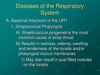
Obstructive and Restrictive Pulmonary Diseases 1. Obstructive disease: characterized by limitation of airflow owing to partial or complete obstruction at any level from trachea to respiratory bronchioles. Pulmonary function test: limitation of maximal airflow rate during forced expiration (FEVI). 2. Restrictive disease: characterized by reduced expansion of lung parenchyma with decreased total lung capacity while the expiratory flow rate is near normal. Occur in: 1. Chest wall disorder. 2. Acute or chronic, interstitial and infiltrative diseases, e.g. ARDS and pneumoconiosis. • Diffuse pulmonary diseases are divided into:
Obstructive disease Restrictive disease
Pathology of lung diseases • Very important in clinical medicine • Complication of air pollution • Common symptoms: • Dyspnea: difficulty with breathing • Decrease compliance, fibrosis • Increased airway resistance , ch. bronchitis • Chest wall disease: kyphoscoliosis, obesity • Fluid accumulation, left sided heart failure • Cough • Postnasal discharge, GERD, Br. Asthma, ch. Bronchitis, pneumonia, bronchiectasis, drug induced • Hemoptysis • Ch. Bronchitis, pneumonia, TB, bronchiectasis, aspergilloma
Atelectasis (collapse) • Incomplete expansion of the lungs or collapse of previously inflated lung substance. • Significant atelectasis reduce oxygenation and predispose to infection.
Types of Atelectasis • Resorptionatelectasis. • Compression atelectasis. • Contraction atelectasis.
Atelectasis • Atelectatic lung is prone to develop superimposed infection. • It is reversible disorder except for contraction atelectasis. • It should be treated promptly to prevent hypoxemia.
Chronic Obstructive Pulmonary Disease Emphysema Bronchiectasis Chronic Obstructive Pulmonary Disease Chronic Bronchitis Asthma
Bronchial asthma • Chronic relapsing inflammatory disorder characterized by hyperactive airways leading to episodic, reversible bronchoconstriction owing to increased responsiveness of the tracheobronchialtree to various stimuli. • triad of: • intermittent and reversible airway obstruction • chronic bronchial inflammation with eosinophils • bronchial smooth muscle cell hypertrophy and hyperreactivity
Bronchial asthma • triggered by environmental antigens, such as: • dusts • Pollen • animal dander • Foods • potentially any antigen is implicated • Drugs, aspirin • sulfur dioxide, ozone, and nitrogen dioxide
Bronchial asthma • It has been divided into two basic types: 1. Extrinsic asthma. 2. Intrinsic asthma.
CLASSIFICATION OF ASTHMA Extrinsic Asthma 70% Intrinsic Asthma 30% • Initiated by diverse, non-immune mechanisms, including: • ingestion of aspirin • pulmonary infections, • cold • inhaled irritant • stress • exercise. • No personal or family history of allergic reaction. • Develop later in life • Initiated by type 1 hypersensivity reaction induced by exposure to extrinsic antigen. • Subtypes include: a. atopic (allergic) asthma. b. occupational asthma. c. allergic bronchopulmonaryaspergillosis. • Develop early in life
Extrinsic Asthma • Atopic (allergic) asthma is the most common form, begins in childhood • Other allergic manifestation: allergic rhinitis, urticaria, eczema. • Skin test with antigen result in an immediate wheel and flare reaction • Other family member is also affected • Serum IgE and eosinophil are increased • immune related, TH2 subset of CD4+ T cells
Pathogenesis of Bronchial Asthma EXAGGERATED BROCHOCONTRICTION • Two components: 1. Chronic airway inflammation. 2. Bronchial hyperresponsiveness. • The mechanisms have been best studied in atopic asthma.
Pathogenesis of Atopic Asthma • A classic example of type 1 IgE-mediated hypersensitivity reaction. • In the airway – initial sensitization to antigen (allergen) with stimulation of TH2 type T cells and production of cytokines (IL-4, IL- 5, and IL-13). • Cytokines promote: 1. IgE production by B cell. 2. Growth of mast cells. 3. Growth and activation of eosinophils.
Pathogenesis of Atopic Asthma • IgE-mediated reaction to inhaled allergens elicits: 1. acute response (within minutes) 2. a late phase reaction (after 4-8 hours)
Pathogenesis of Atopic Asthma Acute-phase response • Begin 30 to 60 minutes after inhalation of antigen. • Mast cells on the mucosal surface are activated. • Mediator produced are : • Leukotrienes C4, D4 & E4 (induce bronchospasm, vascular permeability & mucous production) • Prostaglandins D2, E2, F2 (induce bronchospasm and vasodilatation) • Histamine ( induce bronchospasm and increased vascular permeability) • Platelet-activating factor (cause aggregation of platelets and release of histamine) • Mast cell tryptase (inactivate normal bronchodilator). • Mediators induce bronchospasm, vascular permeability & mucous production.
Pathogenesis of Atopic Asthma • Late phase reaction: • recruitment of leukocytes mediated by product of mast cells including: 1. Eosinophil and neutophilchemotactic factors 2 . IL-4 & IL-5 and induceTH2 subset ofCD4+ T cells 3. Platelet-activating factor 4. Tumor necrosis factor. • Other cell types are involved: activated epithelial cells, macrophages and smooth muscle.
Pathogenesis of Atopic Asthma Late phase reaction: • The arrival of leukocytes at the site of mast cell degranulationlead to: 1. Release of more mediators to activate more mast cells 2. Cause epithelial cell damage . • Eosinophils produce major basic protein, eosinophilic cationic protein and eosinophilperoxidase ( toxic to epithelial cells). • These amplify and sustains injury without additional antigen.
Non-Atopic Asthma • Triggered by respiratory tract infection including viruses and inhaled air pollutants e.g. sulfur dioxide, ozone. • Positive family history is uncommon. • Serum IgE – normal. • No other associated allergies. • Skin test – negative. • Hyperirritability of bronchial tree. • Subtypes: 1. Drug-induced asthma. 2. Occupational asthma.
Morphology of Asthma • Grossly: - lung over distended (over inflation), occlusion of bronchi and bronchioles by thick mucous. • Histologic finding: • Thick BM. • Edema and inflammatory infiltrate in bronchial wall. • mucous contain Curschmann spirals, eosinophil and Charcot-Leyden crystals. • Submucosal glands increased. • Hypertrophy of the bronchial wall muscle.
Curschmann spirals • Coiled, basophilic plugs of mucus formed in the lower airways and found in sputum and tracheal washings
Charcot-Leyden crystals. • Eosinophilic needle-shaped crystalline structures.
Clinical Coarse • Classic asthmatic attack – dyspnea, cough, difficult expiration, progressive hyperinflation of lung and mucous plug in bronchi. This may resolve spontaneously or with Rx. • Status asthmaticus – severe cyanosis and persistent dyspnea, may be fatal • Superimposed bacterial infection • May progress to emphysema or chronic bronchitis • Asthmatic bronchitis: chronic bronchitis with superimposed asthma
Chronic Obstructive Pulmonary Disease Emphysema Bronchiectasis Chronic Obstructive Pulmonary Disease Chronic Bronchitis Asthma
Bronchiectasis • Chronic necrotizing infection of the bronchi and bronchioles leading to or associated with abnormal dilatation of these airways. • Bronchial dilatation should be permanent.
Conditions associated with Bronchiectasis Localized: - tumor, foreign bodies or mucous impaction Generalized: - bronchial asthma - chronic bronchitis • Bronchial obstruction • Congenital or hereditary conditions: • Necrotizing pneumonia - Congenital bronchiactasis - Cystic fibrosis. - Intralobar sequestration of the lung. - Immunodeficiency status. - Immotile cilia and kartagner syndrome Caused by TB, staphylococci or mixed infection.
Kartagener Syndrome • Inherited as autosomal recessive trait. • Patient develop bronchiactasis, sinusitis and situs invertus. • Defect in ciliary motility due to absent or irregular dynein arms. • Lack of ciliary activity interferers with bronchial clearance. • Males have infertility.
Bronchiectasis Etiology and pathogenesis • Obstruction and infection. Bronchial obstruction (athelectasis of airway distal to obstruction) – bronchial wall inflammation. • These changes become irreversible: 1. If obstruction persist. 2. If there is added infection.
Morphology of Bronchiectasis • Usually affects lower lobes bilaterally (vertical airways). • Dilated airways up to four times of normal, reaching the pleura. • Acute and chronic inflammation, extensive ulceration of lining epithelium with fibrosis.
Bronchiectasis • Clinical course: • Sever persistent cough with sputum (mucopurulent, fetid sputum) sometime with with blood. • Clubbing of fingers. • If sever, obstructive pulmonary function develop. • Rare complications: metastatic brain abscess and amyloidosis.
Summary • Asthma • Bronchiectasis
Bronchiectasis: Chronic necrotizing infection of the bronchi and bronchioles leading to permenant dilatation of these airways
Questions • What triggers an attack of asthma?