Download

1 / 69
700 likes | 714 Views
Approach to sellar mass and pituitary stalk lesions. Majid Valizadeh Research institute for endocrine sciences. Outline:. Sellar and parasellar mass different pathology Presentation Approach to Sellar mass Stalk lesions different pathology Presentation

E N D
Approach to sellar mass and pituitary stalk lesions Majid Valizadeh Research institute for endocrine sciences
Outline: • Sellar and parasellar mass • different pathology • Presentation • Approach to Sellar mass • Stalk lesions • different pathology • Presentation • Approach to Stalk lesions • Take home message
Introduction: • the pituitary gland, (hypophysis )is an endocrine gland about the size of a pea and weighing 0.5 grams in humans. • Anterior • Intermediate • Posterior
Importance: • Theprevalence of pituitary adenomas per 100,000 was fourfold higher than previous estimates : • ●All adenomas – 77.6 • ●Lactotroph adenomas – 44.4 • ●Nonfunctioning adenomas – 22.2 • ●Somatotroph adenomas – 8.6 • ●Corticotroph adenomas – 1.2
Classification: • Size: • Microadenoma • Macroadenoma • Cell of origin: • Gonadotroph adenomas • Thyrotroph adenomas • Corticotroph adenomas • Lactotroph adenomas • Somatotroph adenomas • plurihormonaladenomas: // Lactotroph/somatotroph adenoma combinations • Location: • Intrasellar • Extrasellar • Mixed
Presentations of sellar and parasellar mass • ● Neurologic symptoms, such as visual impairment or headache • ● Incidental finding on MRI performed for some other reason • ● Hormonal abnormalities (under- or oversecretion of pituitary hormones)
Causes: • Pituitary adenomas are the most common cause of sellarmasses (about 90%) • Other disorders, which are often difficult to distinguish from pituitary adenomas by imaging, include physiologic enlargement of the pituitary and benign and malignant tumors • Pituitary hyperplasia: • Lactotroph hyperplasia • Thyrotroph and gonadotroph hyperplasia • Somatotroph hyperplasia • Other benign tumors • Craniopharyngiomas, meningiomas, and, less commonly, pituicytomas
Causes: • Malignant tumors: • Primary: germ cell tumors (ectopic pinealomas), sarcomas, chordomas, lymphomas and rarely Pituitary carcinomas. • Metastatic disease — Metastases to the hypothalamus and pituitary gland account for 1 to 2% of sellar masses (most commonly: breast cancer in women / lung cancer in men) • Cysts — Several types of cysts can occur in the sellar and/or suprasellar area, including Rathke's cleft , arachnoid, and dermoidcysts • Abscess: only one third have features suggestive of infection (fever, leukocytosis, meningismus) • Arteriovenous fistula of the cavernous sinus • Hypophysitis:usually occurs in late pregnancy or the postpartum period
How distinguish • Why: Correct preoperative diagnosis is clinically important because the treatment of choice for many of these nonpituitarysellar masses differs from that of a pituitary tumor. (Germ cell tumors, Sarcoidosis, LCH, Lymphoma ) • In some cases, there are no features that clearly distinguish the unusual etiologies from the clinically nonfunctioning pituitary adenoma. • In others, certain endocrine, neurologic, and radiographic findings that are more characteristic of patients with a nonpituitarysellarmass may be present and can help in their differentiation.
Evaluation of sellar mass: • Should be evaluated both radiologically and hormonally. (Clinically- Visual field) • Certain MRI findings: suggest a greater likelihood of some kinds of sellarmasses As an example, a mass that is separate from the pituitary gland generally indicates that the mass is not a pituitary adenoma. • However, no finding is usually pathognomonic of any one kind of mass
SIGNS AND SYMPTOMS OF NONPITUITARY SELLAR LESIONS • Clinical DI at presentation is highly suggestive of a nonpituitary etiology of a sellar or parasellarmass. Sarcoidosis and metastatic disease to the sellar region are especially likely to lead to diabetes insipidus. • The (SIADH) syndrome of inappropriate antidiuretic hormone secretion leading to • potentially severe hyponatremia may also occur in patients with nonpituitary • sellar and parasellar lesions.
SIGNS AND SYMPTOMS OF NONPITUITARY SELLAR LESIONS • Hypothalamic tumors in children may produce the diencephalic syndrome manifest as wasting, poor development, and sexual immaturity. • Hypothalamic dysfunction in adults may lead to disruption of the control of appetite and cause syndromes of polyphagia and massive obesity or severe starvation.
MRI: • Unenhanced image The normal anterior pituitary gland demonstrates a signal intensity similar to or slightly greater than white matter, whereas the posterior pituitary gland appears bright in most patients owing to the presence of phospholipid within the neurosecretory granules. This bright spot may not be seen in 10% to 20% of normal individual. • Pituitary adenomas, by contrast, are most often hypointense relative to the normal gland on T1-weighted images, and approximately one third to one half are hyperintense on T2- weighted images. • n delayed images, this pattern may be reversed. • Pituitary adenomas may be confined to the sella, extend suprasellarly toward the optic the lack of sellar enlargement is helpful in diagnosis, which is suggestive for a nonpituitary lesion.
Gadolinium-enhanced image • Normal pituitary tissue takes up gadolinium to a greater degree than CNS tissue and therefore has a higher-intensity signal than the surrounding CNS. Both micro- and macroadenomas of the pituitary (as well as other sellar masses such as craniopharyngiomas and meningiomas) usually take up gadolinium to a lesser degree than the normal pituitary but more than the CNS. • Therefore, the degree of gadolinium enhancement does not distinguish one kind of sellar mass from another. The postcontrast enhancement of meningiomas is usually homogeneous. • If a sellar lesion can be seen as separate from the normal pituitary, whether on unenhanced or, more commonly, enhanced images, the lesion is not a pituitary adenoma
Some lesions: • Cystic lesions, such as Rathke's cleft cysts: • On T1-weighted images low-intensity signal; however, craniopharyngiomas and even pituitary adenomas may be partially cystic and, therefore, have low-intensity signals. Furthermore, the signal intensity on T1-weighted images will be high if the protein or lipid concentration of the cyst fluid is high. • On T2-weighted images, cystic lesions may have a high-intensity signal . • Hemorrhage into the pituitary gland results in a high-intensity signal on both T1- and T2-weighted images. • Meningiomas typically have a brighter and more homogeneous signal than pituitary adenomas. They also have a suprasellar rather than a sellar epicenter and a dural-based attachment best seen after contrast enhancement
CT scan: • Calcification in a craniopharyngioma or meningioma is seen better by CT scan than by MRI.
Visual loss • The particular visual field loss may provide some clue as to the nature of the lesion. • Unilateral visual loss: Lesions anterior to the chiasm, such as meningiomasof the optic nerve sheath • homonymous hemianopsias: lesions compressing the visual system more posteriorly along the optic tract, such as meningiomas or aneurysms. • bitemporal field cuts of the classic superior chiasmal compression variety. • more unusual visual deficits: Lesions involving the chiasm, such as gliomas • Cranial neuropathy: nonpituitary masses more commonly originate from or infiltrate parasellarstructures
Visual loss pattern: • Unilateral visual loss • homonymous hemianopsias • bitemporal field cuts • more unusual visual deficits
Hormonal evaluation • Hormonal hyposecretion— Usually has no value in the differential diagnosis of a sellar mass. • Can be caused by any hypothalamic or pituitary lesion • One exception to this statement is that the spontaneous development of central DI indicates that the lesion affects the hypothalamus or the stalk and is therefore not a pituitary lesion. • The possibility of hormonal hyposecretion should be evaluated in all patients who have a sellar mass in order to identify and replace hormone deficiencies.
hypersecretion • Acromegaly • Hyperprolactinemia • Cushing and Subclinical Cushing
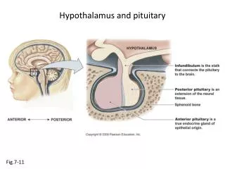
Anatomy • The normal pituitary stalk is widest superiorly and tapers inferiorly. • near the median eminence:3.5 mm, • at its midpoint: 2.88 mm, • at its insertion at the pituitary: 1.9mm • Enlargement of the pituitary stalk greater than 2–3 mm in MRI is pathologic • On MRI: • T1-weighted images, the signal intensity of the stalk is less than that of the optic chiasm and neurohypophysis. • Deviation or tilt of the pituitary stalk can be seen without any underlying abnormality
Pituitary stalk: • Within the pituitary stalk are the axons carrying vasopressin and oxytocin • within the stalk are the pituitary portal vessels that transport the various releasing and inhibiting factors collected in the venous plexus of the median eminence to the pituitary sinusoids • As a consequence of this critical position of the pituitary stalk, patients who develop • pathology involving the stalk commonly present with varying degrees of hypopituitarism, diabetes insipidus, and hyperprolactinemia.
Pituitary stalk lesions • Discovered on MRI: • incidentally • to investigate symptoms such as DI • Classification: • Congenital and developmental • Inflammatory and infectious • Neoplastic
Congenital lesions • pituitary hypoplasia: • Clinically, patients present with short stature because of GH deficiency • On MRI, these patients can • have a hypoplasic, absent, or a short, thickened stalk and an ectopic posterior pituitary
septooptic dysplasia • midline forebrain abnormalities, optic nerve hypoplasia, and hypopituitarism. • On an MRI of a patient with septo-optic dysplasia, one may see lack of • an infundibulum, anterior pituitary hypoplasia, and an ectopic or undescended posterior pituitary. • Clinically, GH deficiency is seen first, • mutation in the pituitary transcription • factor HESX1
Duplication of the pituitary • These cases are often associated with midline facial abnormalities and many of these patients die in infancy
Inflammatory and infectious lesions • Infundibuloneurohypophysitis(LINH) • the inflammation is limited to the infundibulum and posterior lobe, the term lymphocytic infundibuloneurohypophysitis (LINH) • the most common inflammatory cause of pituitary stalk abnormalities • MRI: thickening of the pituitary stalk, loss of the tapering at the pituitary insertion, and marked enhancement with gadolinium, the normal enhancement of the neurohypophysis is absent on MRI, and clinically DI is present
infundibuloneurohypophysitis (LINH): • If the adenohypophsis (infundibulopanhypophysitis)is also involved, anterior pituitary deficiencies can occur. • Corticotropin(ACTH) is the most common anterior pituitary hormone affected, followed by thyrotropin, gonadotropins, and prolactin. • Pathology: Lymphocytic Infiltration • In LINH, the inflammation can be self limited and regression of the lesion can be seen on follow up imaging. / DI, however, tends to be permanent. • In contrast to lymphocytic hypophysitis: a male predominance and the mean age of occurrence is 47 years. • The diagnosis of LINH is made based upon clinical, laboratory, and imaging findings; however, a definitive diagnosis can only be made with biopsy.
LINH treatment: • Glucocorticoids can be used to treat LINH. • the response to glucocorticoids was more pronounced in those with disease duration < 6 months. • Improvement in MRI findings occurred in the majority of patients within 6 weeks to 6 months of treatment.
Langerhans cell histiocytosis (LCH) • Involves the skin, bones, orbit, lungs, and CNS • Granulomas are formed from a proliferation of histiocytes • MRI: asymmetrically thickened pituitary stalk or a hypothalamic • Mass that is isointense on T1 images, hyperintense on T2 images, and enhances with gadolinium • Loss of posterior pituitary enhancement
Diagnosis and Treatment • Search for extracranial manifestations of LCH with a radiographic skeletal survey, skull series, chest radiograph, and bone scan so that these lesions can be Biopsied. • Local radiotherapy (1000–2500 cGy) alone or with chemotherapy (etoposide, vinblastine, and/or cyclosporine).
sarcoidosis • Neurosarcoidosis 5-15% - Usually in context of widespread disease • DI occurs in 25% of patients with CNS sarcoid. • MRI: pituitary stalk thickening and enhancement as well as pituitary enlargement. Periventricular lesions and leptomeningeal enhancement can be seen in sarcoidosis and this can help distinguish it from lymphocytic hypophysitis. • A chest radiograph, (CSF) angiotensin converting enzyme (ACE),
Wegener’s granulomatosis • Systemic vasculitis that causes necrotizing granulomas in the upper and lower respiratory tracts and kidneys. • mean age of onset is 40 and there is a 2 : 1 male to female. • Involvement of the pituitary can occur: • via direct extension from nasal, paranasal, or orbital disease, • from remote granulomatous involvement • from vasculitis of the hypothalamus • Clinically, patients most frequently have DI but hyperprolactinemia and panhypopituitarism have also been reported.
Wegener’s granulomatosis • MRI: an enlarged pituitary with homogenous enhancement as well as thickening and enhancement of the pituitary stalk, and enhancement of • the optic chiasm • Wegener’s granulomatosis can be treated with glucocorticoids and/or cyclophosphamide.
Craniopharyngioma • have a bimodal peak of incidence: • occurring predominantly in children between the ages of 5 and 10 years, • second smaller peak in incidence occurs in the sixth decade. • There is a female preponderance. • Most craniopharyngiomaspresent as a calcific, cystic suprasellarmass.
Craniopharyngioma • The solid portions typically appear isointense or hypointense on T1-weighted images and hyperintenseon T2-weighted imagesa1 but can also have a mottled appearance owing to calcific regions on MR imaging. • Cystic components demonstrate a high signal on T1- weighted images owing to their high protein content or hemorrhagic components. • Tumoral calcification, which may be best appreciated on CT scan, • Dl is particularly common and is seen in 70% to 90% of childhood craniopharyngiomas • and 40% to 60% of adulthood tumors.
Neoplasms • Germinomas typically present as a hypothalamic or pineal mass, however, they can also manifest as isolated pituitary stalk thickening. • first two decades of life and both sexes are affected equally. • (hCG) or alpha-fetoprotein (aFP) • The clinical manifestations of suprasellargerminomas: include DI, hypopituitarism, and vision changes
Germinomas • germinomas progress within 1.3 years of the discovery of pituitary stalk thickening, and within 2.5 years of the diagnosis of DI. • PET scan can be used to assist in making the diagnosis. The PET scan • will be positive if the patient has a germ cell tumor, and can help distinguish from processes such as histiocytosis. • germinomas are highly radiosensitive • Adjuvant chemotherapy can be given to reduce the radiotherapy doses
Metastases to the infundibulum • The most common malignancies that • result in pituitary metastases are breast and lung cancers • typically occur in older patients and • are often locally invasive and have rapid growth.
Primary tumors • that can involve the pituitary stalk include gliomas such as: • astrocytomas, ependymomas, and pleomorphic xanthoastrocytomas • Pituicytomas (also called infundibulomas)
Investigating a pituitary stalk lesion • Preliminary investigations: • • Anterior pituitary hormones (FSH , LH, testosterone (men), oestradiol(women), ACTH, cortisol, TSH, fT4, prolactin, GH, IGF-1) • • Full blood examination, blood film, biochemistry, calcium and phosphate, renal and liver function • • Markers of cell turnover (LDH, B2M) and inflammation • CRP (C-reactive protein), ESR (erythrocyte sedimentation rate) • Serum ACE, 1,25 dihydroxyvitamin D • • Serum AFP (alpha-fetoprotein) and hCG (human chorionic • gonadotropin) • • c-ANCA (cytoplasmic antineutrophil cytoplasmic antibody) • Quantiferon Gold in the presence of risk factors for tuberculosis, such as travel within an endemic area and immunocompromised states. • Urinalysis • • Urine microscopy and culture • • 24 h urine calcium excretion • Imaging • • CT neck, chest, abdomen and pelvis with contrast
Incidental pituitary stalk lesion Symptomatic pituitary stalk lesion Tumour markers of solid organ malignancy • For selected patients in whom metastatic malignancy is suspected. • e.g. CEA, Ca125, Ca199, PSA. Serum IgG4 • For patients with features suggestive of IgG4-related hypophysitis, such as autoimmune pancreatitis and chronic sinusitis. CSF analysis • CSF analysis for cytology, flow cytometry, immunohistochemistry and polymerase chain reaction (PCR) may be diagnostic in suspected CNS lymphoma and obviate the need for intracranial tissue biopsy. • clinical review and repeat pituitary hormone testing in 3 months, and MRI pituitary in 6 months, and thereafter every 6–12 months during the first 2– 3 years. • If the PS lesion increases significantly in size (>6.5 mm in width) or new lesions develop, or if pituitary hormone deficiency arises or the patient’s clinical status changes, then we suggest proceeding to more invasive investigations including consideration of PS biopsy.
Symptomatic pituitary stalk lesion Bone marrow aspirate and trephine (BMAT) • For patients with suspected CNS lymphoma as a confirmatory and staging investigation. Tissue biopsy • Histopathology is required for a diagnosis of metastatic disease from solid organ malignancy, neurosarcoidosis, CNS lymphoma, GCT and LCH. • If peripheral tissue is not available, then PS biopsy should be considered. • • CSF analysis for microscopy and culture is indicated in patients with risk factors, or clinical or radiographic features suggestive of CNS tuberculosis. • • CSF analysis for AFP and hCG should be performed in patients with radiographic features of GCT in whom serum AFP and hCG are negative. • Testicular ultrasongraphy • • Should be performed in men with features of CNS lymphoma • to exclude occult testicular lymphoma. • Other imaging modalities • • In suspected cases of LCH, radiographic skeletal survey, • chest X-ray and whole body bone scan are indicated to assess • for extracranial granulomas amenable to biopsy.