Download
1 / 18
220 likes | 583 Views
Hypothalamus and pituitary. Fig.7-11. Posterior pituitary. HYPOTHALAMUS. POSTERIOR PITUITARY. Vein. Fig.7-12. Anterior pituitary. HYPOTHALAMUS. Neurons. Capillary bed . Artery. Portal vessels. POSTERIOR PITUITARY . Endocrine cells. Capillary bed . ANTERIOR PITUITARY . Veins.

E N D
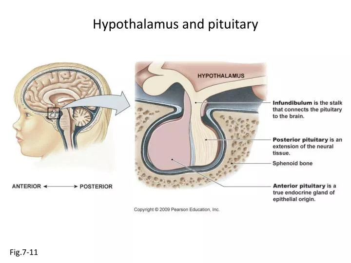
Hypothalamus and pituitary Fig.7-11
Posterior pituitary HYPOTHALAMUS POSTERIOR PITUITARY Vein Fig.7-12
Anterior pituitary HYPOTHALAMUS Neurons Capillary bed Artery Portal vessels POSTERIOR PITUITARY Endocrine cells Capillary bed ANTERIOR PITUITARY Veins TO TARGET ORGANS Fig.7-12
Adrenal gland Adrenalmedulla Adrenalcortex Fig. 23-1
Bone growth Epiphysis Diaphysis Bone growth Compactbone Dividing chondrocytes Chondrocyte Chondrocytes Cartilage Old chondrocytes Epiphyseal plate Osteoblast Direction of growth Diaphysis Osteoblasts Newlycalcifiedbone Fig. 23-16
Acromegaly Fig. 23-14
Functions of muscle Triceps musclecontracts (extensor) Tricepsmusclerelaxes Bicepsmusclerelaxes Biceps musclecontracts (flexor) Flexion Extension Fig. 12-2
Functions of muscle Peristalsis Vasoconstriction Hydrostatic Pressure Fig. 12-2
Types of muscle Nucleus Muscle fiber(cell) Striations Skeletal muscle Fig. 12-1
Types of muscle Striations Muscle fiber Intercalated disk Nucleus Cardiac muscle Fig. 12-1
Types of muscle Muscle fiber Nucleus Smooth muscle Fig. 12-1
Muscle structure Skeletal muscle Nerve andblood vessels T endon Connective tissue Muscle fascicle:bundle of fibers Connectivetissue Nucleus Muscle fiber Fig. 12-3
Muscle structure ULTRASTRUCTURE OF MUSCLE Mitochondria Sarcoplasmicreticulum Thickfilament Thinfilament Nucleus T-tubules Myofibril Sarcolemma Fig. 12-3
Muscle structure Sarcomere Z disk Z disk Myofibril Titin Z disk Z disk Myosin crossbridges M line M line Thick filaments Thin filaments Titin Troponin Myosin heads Hinge region Myosin tail Tropomyosin G-actin molecule Myosin molecule Actin chain Fig. 12-3
Cross-bridge cycle 1 Cytosolic Ca2+ Troponin G-Actin 3 Tropomyosin shifts,exposing bindingsite on actin 2 TN TN 5 Myosin head Actinmoves Tropomyosinblocks bindingsite on actin ADP ADP 4 Pi Power stroke Pi Relaxed state. Myosin head cocked. Initiation of contraction Fig. 12-9
Sarcomere contraction Fig. 12-8