Download
1 / 22
220 likes | 223 Views
Learn about the different cell types and structures, and how they relate to the development of vaccines and treatment methods for infections. Investigate the specific differences between our cells and pathogens, and understand how antibodies and vaccines work.

E N D
Content: Cell Types and Structures • Vaccines are often developed against specific antigens found only in one pathogenic organism. • There are other ways to treat infections of pathogenic organisms, that usually take advantage of many of the other specific differences that exist between our cells and the pathogens. • We will investigate the large differences today and then talk about antibodies and vaccines in the next class.
Learning Objectives • Identify or recall the different structural components and reproductive strategies present in prokaryotes, eukaryotes, and viruses. • Given data about an organism, apply your knowledge of prokaryotes, eukaryotes, and viruses to determine what the organism is. • Explain why various treatment methods work to specifically kill one class of organisms while remaining harmless to human cells or other organisms.
Unknown Infection • Servicemen in Middle East. • Looks like insect bites maybe… • Pretty scary insects though, and what if it’s not an insect? • What else could it be?
Suspect 1 - Virus Herpes: Is spread through skin-skin contact. Symptoms include blisters on the skin or mucous membranes (chicken pox, shingles, cold sores, genital herpes, mono). The virus has a double strand of DNA (74 genes) surrounded by protein cage (capsid) and phospholipidbilayer (180-200nm Source: wikimedia images
Suspect 1 : Virus Size • Smallest (50nm) • 100 times smaller than bacteria Composition • Outer envelope: repetitive protein often inserted into a lipid membrane (responsible for recognition and infection of host cell). • Protected capsid that contains genetic material (DNA or RNA) with important protein enzymes required for duplication. Cannot reproduce by itself • hijacks a host cell to replicate itself. Source: wikimedia images
Virus hijacking host system Many antiviral drugs work by blocking specific enzymes used by the virus for duplication or infection of host cells. Acyclovir, the most common drug used for Herpes infections, affects the viral protein that duplicates DNA.
Clicker Question 1 As obligate intracellular parasites, viruses have which of the following characteristics: After entering a cell, they manufacture their own ATP and carbon-containing compounds like proteins and nucleic acids in order to survive. After entering a cell, they use the host cell's machinery to make more copies of themselves using host proteins. After entering a cell, they use their own protein-synthesizing machinery to make more viral proteins that are used to assemble more copies of themselves.
Suspect 2:S. aureus Bacteria Staphylococcus aureus are 0.6-1.5 µm gram-positive bacterium that have become increasingly drug resistant (MRSA, methicillin resistant S. aureus) It was first discovered in 1880 by a Scottish surgeon in pus from surgical abscesses. Half a million people in American hospitals contract Staph infections each year. Source: wikimedia images
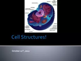
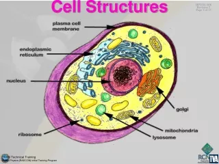
Prokaryotes • Unicellular • Reproduce asexually • Composition • Protected interior (cytoplasm) that contains genetic material (one circle of DNA) as well as protein enzymes to carry out necessary functions of gathering energy, manufacturing proteins (ribosomes), etc…
Prokaryotes • Size • 0.2-10 micrometer (µm) • Composition • A bacterial cell is typically surrounded by a plasma membrane & cell wall containing peptidoglycan (carbohydrates + amino acids). • The amount of peptidoglycan determines differences in their staining properties. Two major categories of bacteria: • Gram positive: Have large amounts of peptldoglycan and stain with a Gram stain. • Gram negative: Have small amounts of peptidoglycan and do not stain with a Gram stain. Almost all Gram negative bacteria are pathogens.
Clicker Question 2 Suppose that a patient contracts a Staph infection.Using Table 1 information, which describes its expected characteristics?
Clicker Question 3 Based on what we know about S. aureus, rank order the antibiotics from best choice to worst choice. Be prepared to provide a rationale for your choice. • Amoxicillin, Penicillin, and other ß-lactams- Blocks the enzyme that normally creates links in peptidoglycan molecules. Kill Gram positive bacteria. 2) Streptomycin - Blocks prokaryotic ribosomes. Effective on many Gram-negative and some Gram-positive bacteria. 3) Ciprofloxacin hydrochloride (Cipro) - Blocks bacterial enzyme needed to prepare DNA for copying. Effective on Gram-negative and Gram-positive bacteria. All antibiotics are equally good choices. 1, 2, 3 3, 2, 1 3 or 2, 1 E. Neither Becky (nor Ellie) should take antibiotics at this point.
Microscope Analysis “Clinical examination and staining and/or culturing of a specimen of pus or exudate are often adequate for diagnosis. Ultraviolet light (Wood's lamp) is helpful in diagnosing erythrasma and some toe web and fungal infections. Microscopic examination of a KOH preparation of skin scales, nail scrapings, or loose hair is useful for fungal infections. For viral infections, stained smears of vesicle fluid are examined under the microscope for typical cytopathology.” http://www.ncbi.nlm.nih.gov/books/NBK8301/
Microscope Analysis On the left are the pathogens infecting the servicemen. On the right is a light microscope photograph of a gram stain showing dozens of S aureus. 1μm S aureus (gram stain) Cells infecting Servicemen
Clicker Question 4 Based on these photos, why would you conclude that the pathogens aren’t bacteria? • They lack cell walls. • The pathogens aren’t the right size and the dark stained material indicates that there are multiple chromosomes. • They are too large and have nuclei. • The dark staining material are mitochondria and they don’t exist in bacteria. • Bacteria have flagella and there is no evidence of them in the photos.
EukaryotesProkaryotes REVIEW
Suspect 3: Fungus Ringworm Dermatophytes of the genera Trichophyton and Microsporum are the most common causative agents of this disease of the skin. A 4-8µm fungus with a defined a cell wall. It is passed as spores from skin-skin contact or on inanimate objects.
Suspect 4: ProtistLeishmaniatropica • 6-10 µm single-celled protozoan parasite of • Transmitted through the bite of a sand fly (Sexual life cycle)
Anti-Eukaryotic Medicines: • Pyrimethamine, Sulfonamides: Interfere with enzymes used to make the folic acid needed to make thymine and uracil nucleotides. Pyrimethamine, Sulfonamides work on protists (don’t affect humans). • Polyenes combine with a component of fungal and some bacterial membranes, disrupt and break them. Inhibits ß-glucan, found in cell walls of fungi
Clicker Question 5 There is a specific blood test that can be used to definitively decide if the pathogen is a protist or a fungus. Which of the following things must this test look for? • A: Presence of DNA • B: Presence of ß-glucan-containing cell walls C: Presence of cellulose D: Presence of peptidoglycan cell walls E: Presence of spores
Problem: • Drugs are not working to cure the infection. • Other servicemen are also infected, and they are seeing a real problem with battle-readiness. • Army Medical College researchers have been brought in…. • Your reading assignment and assessment for the next class deal with how to boost immune system response to pathogens.