Download
1 / 56
560 likes | 566 Views
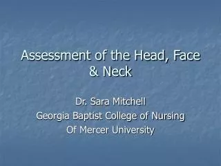
Obstetrical Sonography 1 Lecture 11 HHHoldorf. Fetal Face & Neck. What to take away from this chapter: A fetal profile is standard routine for the OB exam showing the fetal nose, upper and lower lips, chin, and neck. When the fetal orbits are evaluated, the lenses are evaluated.

E N D
Obstetrical Sonography 1 Lecture 11 HHHoldorf Fetal Face & Neck
What to take away from this chapter: • A fetal profile is standard routine for the OB exam showing the fetal nose, upper and lower lips, chin, and neck. • When the fetal orbits are evaluated, the lenses are evaluated. • When measuring the ocular distances, the following may be used Binocular Distance, Inter-ocular distance, ocular diameter. You must refer to the charts to determine if Hyper or Hypotelorism is present. • Facial anomalies associated with holoprosencephaly • Possible forms of cleft lip/cleft palate • Study the 3/D images in this chapter.
Facial Clefting • Cleft lip/palate • Median cleft lip Masses • Epignathus • Teratoma of the neck (cervical teratoma) • Nuchal thickening • Nuchal translucency • Cystic Hygroma
Facial Clefting • The second most common congenital malformation (13% of all fetal anomalies). • Cleft lip/palate • Cleft palate and cleft lip are associated anomalies and are often seen together. Sonographic finding • A groove extending from one nostril through the lip and possibly the alveolar ridge • Usually best demonstrated on coronal section. • Difficult to detect isolated soft palate clefts
Median Cleft Lip • Midline malformation of upper lip with or without cleft palate. Frequently associated with other midline defects of the face and brain, such as Holoprosencephaly. Sonographic findings: • Similar to the diagnosis of cleft lip/cleft palate • Visualization of the tongue in a higher than usual position in the mouth • Again, the face predicts the brain…
MASSESEpignathus • A teratoma arising from the oral cavity or pharynx, and Epignathus may arise from the sphenoid bone, hard or soft palate, pharynx, tongue or jaw. Sonographic features • Solid and cystic complex tumor seen extruding from the fetal mouth • Calcifications may be present within masses
Teratoma of the Neck (Cervical Teratoma) • Similar to Epignathus, except the tumor arises from the neck Sonographic findings • Complex, cystic/solid tumor seen near fetal neck • May mimic a cystic hygroma
Nuchal Thickening • Increased soft tissue thickness over the posterior aspect of the neck. Increased NT is reported to be associated with a slightly higher risk of chromosomal syndromes, especially Trisomy 21. Sonographic findings • Oblique axial cross-section showing CSP, 3rd ventricle, cerebellum and CM • >6 mm considered abnormal, measured between 15 and 21 weeks • Fetal head must not be hyperextended
Cystic Hygroma • Benign developmental anomaly of lymphatic origin characterized by single or multi cystic areas within soft tissues surrounding the neck. • Associated with chromosomal anomalies is common, esp. Turner’s and Down Syndromes. • Large Hygromas are associated with fetal Hydrops. Sonographic findings • Fluid-filled structure presenting as a cystic mass. • May mimic cervical teratoma, NTD
Normal Anatomy Face: • Evaluation of the face is a vital part of the clinical genetic examination that is performed postnatally. Any time a fetal anomaly is identified, the diagnostic workup should include a detailed examination of the fetal face. Many details of facial anatomy can be identified as early as 11 weeks, particularly by using transvaginal sonography. It is necessary that it is accessible and there is a pocket of amniotic fluid in front of it for three dimensional scanning. • The upper lip may be visualized in an oblique coronal plane and is useful in searching for facial clefts and some types of proboscis.
The profile shows the forehead, nose and jaw which are important to asses the integrity of these structures.
Eyes • The eyes may be imaged in either a true coronal or a transverse plane. It is important to measure the outer orbital distance because it is valuable in diagnosing hypertolerism or hypotelorism
Neck • Transverse sections allow the measurement of the nuchal fold. Studies have shown an association with Down syndrome when this measurement exceeds 6mm between 15 and 21 weeks. It is considered nuchal thickening when there is increased soft tissue thickness over the posterior aspect of the neck.
Facial Clefting • Typical facial clefts are cleft lip and Cleft palate anomalies. Cleft palate refers to the defect of the posterior portion of the palate in the presence of a normal upper lip and anterior palate. CL-CP can be unilateral or bilateral, and usually results from a failure of mesenchymal masses of the lateral palatine processes to fuse with each other, with the nasal septum and/ or median palatine process. It can be genetic or non genetic causing a minor developmental defect.
Sonographic presentation demonstrates a groove extending from one nostril through the lip and possibly the alveolar ridge.
A cleft lip or palate can be successfully treated with surgery soon after birth. • The edges of the cleft between the lip and nose are cut (A and B). The bottom of the nostril is formed with suture (C). The upper part of the lip tissue is closed (D), and the stitches are extended down to close the opening entirely (E).
Median cleft lip • A malformation of the upper lip with/without cleft palate. The development of this anomaly is related to the differentiation process of the forebrain and is often associated with other midline defects or the face and brain such as holoprosencephaly. The incidence is about 1: 10,000,000 births • It can vary from being a small notch to a complete division of the lip and alveolar part of the maxilla. • When it is unilateral it results from failure of maxillary prominence to fuse with medial nasal prominences. • When it is bilateral it results from failure of maxillary processes to meet and merge with medial prominences.
Epignathus • A congenital tumor and a rare type of teratoma arising from the oral cavity or pharynx. It is associated with midline abnormalities. It may also arise from the sphenoid bone, hard or soft palate, pharynx, tongue or jaw. • Sonographic Findings: • Solid, complex tumor seen extruding from the fetal mouth, calcifications may be present within mass • Calcifications may be present within mass.
Micrognathism (Micrognathia) • A condition where the jaw is undersized and is sometimes called “Mandibular hypoplasia.” It is common in infants but is usually self correcting during growth, due to the jaws increasing in sized. It may be a cause o abnormal tooth alignment and in severe cases can hamper feeding. It can present as a birth defect in multiple syndromes (fetal alcohol syndrome, congenital rubella, Trisomy 13 and 18)
Macroglossia • Minor or severe enlargement of the tongue can cause cosmetic and or functional difficulties, such as speaking, eating, swallowing and sleeping.
ORBITAL DEFECTS • Hypotelorism- a medical condition pertaining to abnormally close eyes. • Hypertelorism – an abnormally increased distance between two organs or bodily parts, usually referring to an increased distance between the eyes, seen in a variety of syndromes. • Microphthalmia or micropthlamos means small eye.
Anophthalmia (Anopththalmos) • The congenital absence of one or both eyes. • True or primary anopththalmos is very rare. The diagnosis for this can ONLY be made when there is complete absence of the ocular tissue within the orbit. Extreme micropthlamos is seen more commonly. In this condition, a very small globe is present within the orbital soft tissue, which is not visible on the first examination.
Primary (No eye tissue) Secondary (extremely tiny eyes, eyes start to develop then stops) • Degenerative (eye started to form and for some reason degenerated
Retinoblastoma • Cancer of the retina. This tumor can begin in one or both eyes and can spread to the brain through the optic nerve. Development of the tumor is initiated by mutations
Holoprosencephaly • A spectrum of defects or malformations of the brain and face. The most severe cases involve serious malformations of the brain, and malformations so severe that they often cause miscarriage or stillbirth. There can also be facial defects – which may affect the eyes, nose, and upper lip- and normal or near- normal brain development. Seizures and mental retardation may occur. The etiology is unknown. Suggested risk factors include: maternal diabetes, infections during pregnancy such as herpes, syphilis, rubella etc., and various drugs taken during pregnancy. Women with previous pregnancy loss and 1st trimester bleeding are likely to give birth to a child with this defect. Symptoms: • Range from mild (no facial/organ defects), moderate ( cleft lip or palate) to severe ( synopththalmia proboscis or cyclopia)
Holoprosencephaly • The alobar type, which is the most severe, is characterized by a monoventricular cavity and fusion of the thalami. In the semilobar type there is partial segmentation of the ventricles and cerebral hemispheres posteriorly with incomplete fusion of the thalami. In lobar holoprosencephaly there is normal separation of the ventricles and thalami but absence of the septum pellucidum.
Anomalies of the Neck • Teratoma of the Neck (Cervical teratoma) • Similar to epignathus except the tumor arises from the neck. Sonographic appearance includes complex, cystic/solid tumor seen near the fetal neck. • It is vital to be able to identify the origin of the mass in order to distinguish it from epignathus.
Cystic Hygroma • Benign developmental anomaly of lymphatic origin characterized by sing or multiple cystic areas within soft tissues surrounding the neck. Sonographically it can appear to be a thin walled, multi-septated cyst usually located posterior to fetal head/neck but also may be anterior.
Key points in Chapter 11, the fetal face and neck • Hypotelorismis a medical condition pertaining to abnormally close eyes. • Hypertelorism is an abnormally increased distance between two organs or bodily parts, usually referring to an increased distance between the eyes (orbital hypertelorism), seen in a variety of syndromes
Holoprosencephaly • Symptoms of holoprosencephaly range from mild (no facial/organ defects) to moderate (cleft lip or palate) to severe (syn-ophthalmia proboscis or cyclopia).
There are three classifications of holoprosencephaly. • Alobar holoprosencephaly, the most serious form in which the brain fails to separate, is usually associated with severe facial anomalies. • Semilobar holoprosencephaly, in which the brain's hemispheres have a slight tendency to separate, is an intermediate form of the disease. • Lobar holoprosencephaly, in which there is considerable evidence of separate brain hemispheres, is the least severe form. In some cases of lobar holoprosencephaly, the patient's brain may be nearly normal. • Homework: Show the three classifications of Holoprosencephaly.
Holoprosencephaly, consists of a spectrum of defects or malformations of the brain and face. At the most severe end of this spectrum are cases involving serious malformations of the brain, malformations so severe that they often cause miscarriage or stillbirth. At the other end of the spectrum are individuals with facial defects - which may affect the eyes, nose, and upper lip - and normal or near-normal brain development. Seizures and mental retardation may occur.
The most severe of the facial defects (or anomalies) is cyclopia, an abnormality characterized by the development of a single eye, located in the area normally occupied by the root of the nose, and a missing nose or a nose in the form of a proboscis (a tubular appendage) located above the eye. The condition is also referred to as cyclocephaly or synophthalmia and is very rare.
The cause of holoprosencephaly is currently unknown. Often, no specific cause can be identified. Suggested risk factors include maternal diabetes, infections during pregnancy (syphilis, toxoplasmosis, rubella, herpes), and various drugs taken during pregnancy (alcohol, aspirin, lithium, thorazine, anticonvulsants, hormones). Women with previous pregnancy loss and first trimester bleeding are also more likely to have a child diagnosed with holoprosencephaly. • Although many children with holoprosencephaly have normal chromosomes, specific chromosomal abnormalities have been identified in some patients.
Holoprosencephaly is not a condition in which the brain deteriorates over time. Although serious seizure disorders, autonomic dysfunction, complicated endocrine disorders and other life-threatening conditions may sometimes be associated with HPE, the mere presence of HPE does not mean that these serious problems will occur or develop over time without any previous indication or warning. These abnormalities are usually recognized shortly after birth or early in life and only occur if areas of the brain controlling those functions are fused, malformed or absent. • Prognosis is dependent upon the degree of fusion and malformation of the brain, as well as other health complications that may be present.
The more severe forms of holoprosencephaly are usually fatal. This disorder consists of a spectrum of defects, malformations and associated abnormalities. Disability is based upon the degree in which the brain is affected. Moderate to severe defects may cause mental retardation, endocrine disorders, epilepsy and other serious conditions. Whereas, mild brain defects may only cause learning or behavior problems with few motor impairments. • Seizures may develop over time with the highest risk before 2 years of age and the onset of puberty. Most are managed with one medication or a combination of medications. Typically, seizures that are difficult to control appear soon after birth, requiring more aggressive medication combinations/doses.
Anophthalmia, also known as anophthalmos (Greek: "without eye"), is the congenital absence of one or both eyes.
True or primary anophthalmos is very rare. Only when there is complete absence of the ocular tissue within the orbit can the diagnosis of true anophthalmos be made. Extreme microphthalmos is seen more commonly. In this condition, a very small globe is present within the orbital soft tissue, which is not visible on initial examination.
There are three classifications for anopthalmia: • Primary anophthalmia is a complete absence of eye tissue due to a failure of the part of the brain that forms the eye. • Secondary anophthalmia the eye starts to develop and for some reason stops, leaving the infant with only residual eye tissue or extremely tiny eyes which can only be seen under close examination. • Degenerative anophthalmia the eye started to form and, for some reason, degenerated. One reason for this occurring could be a lack of blood supply to the eye.
Retinoblastoma is a cancer of the retina. Development of this tumor is initiated by mutations. • The tumor may begin in one or both eyes. Retinoblastoma is usually confined to the eye but can spread to the brain via the optic nerve.