Download
1 / 50
500 likes | 569 Views
The Immune System Dr. Ammar ALMOFTI. Phagocytes. Neutrophils. 50 ~ 80 % of all leukocytes. An elevation in the number of neutrophils in blood is used clinically as a determinant of infection. Neutrophils release cytokines. Eosinophils. 1 ~ 4 % of all leukocytes

E N D
Neutrophils • 50 ~ 80 % of all leukocytes. • An elevation in the number of neutrophils in blood is used clinically as a determinant of infection. • Neutrophils release cytokines.
Eosinophils • 1 ~ 4 % of all leukocytes • Release toxic substances against parasites.
Monocytes in blood • 2 ~ 8 % of all leukocytes. • Monocytes in blood can be developed into Macrophages in tissues. • Eg: • Microglia : Nervous tissue • Kupffer cells : Liver • Histiocytes: connective tissue
One Additional phagocytic cell • Dendritic cells: • activate T-cells. • they are formed in bone marrow from hematopoietic stem cells
Basophils • secrete chemical mediators (Histamine…) in inflammation and allergic reaction. • ( < 1% of all leukocytes) • Release toxic substances against larger parasites.
Mast cells • Secrete histamine and other substances. • They are found in skin and mucosal epithelial tissue. • They are formed in bone marrow from hematopoietic stem cells
Lymphocytes • Provide: • Diversity • Specificity • Memory • Distinguish between self nonself • Lymphocytes Types: • B- Lymphocytes • T- Lymphocytes • Null cells
B-Lymphocytes • Contact antigens and develop to plasma cells. • Plasma cells secrete antibodies (Immunoglobulins) that mark Ags only.
T-Lymphocytes • Contact infected, mutant and transplanted cells and develop to cytotoxic T-cells. • They kill abnormal cells by secreting perforines; molecules form pores in the membrane causing burst.
Null cells • No cell membrane component. • Mostly called Natural Killers (NK). • Contact virus infected cells and kill them.
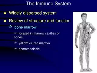
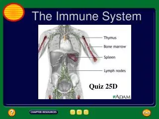
Lymphoid Tissues • Central Lymphoid tissues • Bone marrow. • Thymus • Peripheral lymphoid tissues • Spleen. • Lymph nodes • Tonsils • Adenoids • Appendix • Lymph nodules of the gastrointestinal tract.
Filteration • Spleen: filter blood . • Lymph nodes: filter lymph. • Tonsils and adenoids: filter inhaled particles. • Appendix, lymph nodules and Peyer’s patches: filter substanses in ingested food and water.
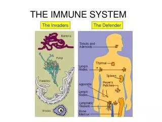
Organization of Body’s Defenses • Nonspecific defenses: • Physical barriers: skin (sebaceous glands, sebum) • Inflammation • Interferons • Complement system • Specific defenses
Inflammation • Phagocytosis by nearby macrophages. • Dilation and permeability of capillary: induced by histamine released by mast cells (basophils) -> redness, swelling, heat, pain. • Containment of foreign matter: • heparin suspends blood clotting temporarily. • Access of leukocytes to injured area. • Clot formation. • Leukocyte migration and proliferation after injury. • Continued clearing of infection by recruited leukocytes.
Cont. • Neutrophils: 1 hour after injury accumulate. • Monocytes: 10 hours after injury accumulate and develop. • Transit regulation of leukocytes: • Margination. • Diapedesis. • Attachment. • Chemotaxis.
Steps of Phagocytosis • Attachment. • Internalization. • Degradation. • Exocytosis.
Opsonization for attachment • Attachment is enhanced by opsonins. • Opsonins are proteins (including antibodies) that bind tightly to the foreign material and make it easier for phagocytosis.
Cytokines secreted by macrophages • Interleukin-1 (IL-1). • Interleukin-6 (IL-6). • Tumor necrosis factor – alpha (TNF- α).
Functions of cytokines • Synthesis of more adhesion molecules by blood vessel endothelial cells. • Release of more neutrophils from bone marrow. • Action on the hypothalamus to raise body temperature by releasing prostaglandins. • Stimulation of liver cells to produce acute phase proteins, antibacterial proteins such as C-reactive protein which acts as an opsonin. • IL-1 helps in proliferation and differentiation of B and T lymphocytes.
Interferons • Interferons are a group of proteins interfere with virus replication. • Interferon-α and Interferon-β are secreted from virus-infected cells and their function is to induce resistance to the surrounding cells. • Interferon-γ is secreted by active T cells and NK cells. Its function is to inhibit viral replication, enhance phagocytosis, boost antibody production, activate NK and cytotoxic T cells, finally suppress growth of tumor.
The Complement System • Its so named because it completes the actions of specific antibodies, but this system acts in the absence of antibodies. • The system consists of about 30 plasma proteins as a cascade of activation steps to destroy invading microorganisms by development of a membrane attack complex (MAC) which forms a pore in bacterial membrane and so bacteria will burst.
Other functions of complement system • Some complement proteins act in chemotaxis, guiding phagocytes into the area. • Others bind to nearby mast cells and induce them to release histamine. • One specific protein, called C3b, coats bacterial surfaces were it acts as an opsonin.
Humoral Immunity • It is a specific immune response generated by B lymphocytes and conferred by antibodies that circulate in the blood and lymph. • B lymphocytes proliferate and develop into plasma cells and memory B cells. The plasma cells secrete antibodies.
Cell-Mediated Immunity • It is the reaction of certain types of T lymphocytes to kill abnormal or infected body cells. • T lymphocytes proliferate and develop into cytotoxic T cells and memory T cells. The cytotoxic T cells kill abnormal or infected body cells.
Primary Immune Response • When a person is first exposed to an antigen. • The B cells and T cells proliferate and develop into effector cells and memory cells (clonal selection). • This reaction takes place about 10-17 days after exposure to the antigen.
Secondary Immune Response • When a person is subsequently exposed to the same antigen. • It is faster; only 2-7 days. Memory cells quickly proliferate and differentiate into effector cells and again memory cells. • Greater in magnitude. • More prolonged than primary response.
Important Note • Helper T cells sometimes influence B cell activation when contact antigens by secreting cytokines (including IL-2). In this case, antigens are called T-dependent antigens. • Some antigens do not need helper T cells. They are called T-independent antigens. In this case, B cells do not develop to memory B cells and so repeated exposures to the same antigen always cause primary response.
Classes of Antibodies • IgG • IgE • IgM • IgD • IgA • All Igs neutralize and agglutinate antigens
IgG • The most common in the blood produced in secondary responses. • Crosses placenta, so it is important for fetus and newborns. • Other functions of IgG: • IgG activates complement. • Opsonizes antigens. • Enhances NK cell activity.
IgE • Involved in allergies • Binds to mast cells and basophils, causing them to release histamine.
IgM • The most common produced in the primary response. • Activates complement.
IgA • It crosses epithelial cells and it is present in breast milk, so it is important in immunity in newborns.
T lymphocytes • Helper T cells. • Cytotoxic T cells. • Suppressor T cells. Suppressor T cells are not well-understood.
Helper T cells • Operate indirectly. • Secrete cytokines to activate B cells, T cells including helper T cells themselves. • Secrete cytokines to activate macrophages and NK cells
Cytotoxic T Cells • Operate directly. • They kill cells infected by viruses. • They kill abnormal cells (like cancer cells).
T cell Receptor (TCRs) • TCRs detect antigen only when antigen is associated with molecules called MHC (Major HistoCompatibility). • MHC molecules first bind to antigen (or fragment) within a cell body and then transport it to the surface to be detected by T cells. This process is called antigen presentation.
MHC molecules • MHC molecules in human are called HLA molecules (human leukocyte antigens). • They are varied enough to various complex antigens but unique to each person. • So, each person has different HLA molecules to recognize a different portion of the antigen. • This diversity of HLA molecules is adaptive to the survival of the human species as a whole.
Classes of MHC molecules • Class I MHC molecules: are found on the surface of all nucleated cells (every cell of the body). • Class II MHC molecules: are found on the surface of some specialized cells as: • macrophages and dendritic cells. • activated B cells.
MHC-I molecules & Antigen Presentation • Class I MHC molecules capture fragments of antigen within an infected cell and transport them to the surface. • A cytotoxic T cell then binds to the infected cell through its TCR and CD8. This cytotoxic T cell is called CD8 cell.
How do CD8 cells kill? • CD8 cells kill virus-infected cells by releasing perforins. • CD8 cells release fragmentins (proteins enter through the pores and cause apoptosis to the infected cells). • CD8 cells kill tumor cells after binding of their TCRs to the distinctive tumor antigens presented by class I molecules existing on tumor cells. • DOES TUMOR INHIBIT (CLASS-I)PRODUCTION?
MHC-II molecules & antigen presentation • Class II MHC molecules capture fragments of antigen engulfed by a macrophage and transport them to the surface. • A Helper T cell then binds to the presenting cell through TCR and CD4. This helper T cell is called CD4 cell. • CD4 cells do not kill the antigen-presenting cell but secrete cytokines to activate other immune cells.
Immunization • It is called also vaccination. • A safe form of a microorganism (or a collection of its components) is introduced into the body and then stimulates immune response and immunological memory. • It could be artificial or natural after real infection.
Active Immunity • Both artificial and natural immunization confer a type of protection referred to as active immunity. • Because it depends on the immune system to mount a response.
Passive Immunity • Ready-made antibodies to a particular antigen can also be introduced into the body to provide a protection called passive immunity. • Can passive immunity be natural? IgG-IgA?
Allergy • It is called hypersensitivity reaction. It is exaggerated response to certain allergens. • IgE is the most involved antibody. • IgE is produced highly and causes hay fever in restricted area when an individual responds to pollen.
Anaphylactic shock • It is a life-threatening reaction to injected or ingested allergens. • Widespread degranulation of mast cels. • Dilation of peripheral blood vessels. • Drop in total peripheral resistance. • Drop in pressure. • Death may occur within a few minutes.
Management of Allergic shock • Injection of epinephrine (adrenaline) raises blood pressure back toward normal levels. END