Download

1 / 21
290 likes | 474 Views
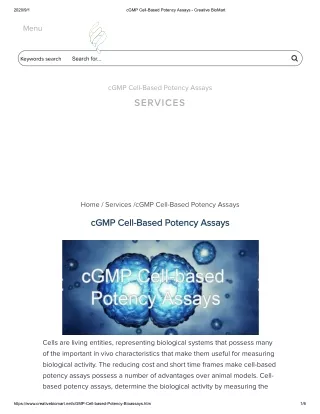
Apoptosis is considered a vital component of various processes including normal cell turnover, proper development and functioning of the immune system, hormone-dependent atrophy, embryonic development and chemical-induced cell death.<br><br>https://www.creative-bioarray.com/cell-apoptosis-assays.htm

E N D
Spotlight on —— APOPTOSIS Creative Bioarray
CONTENTS CreativeBioarray Part 1 Part 2 Part 3 Part 4 Introduction Disease Detection Mechanism — 2—
Introduction Part 1 — 3— Apoptosis is a form of programmed cell death, or “cellular suicide.” Apoptosis is an orderly process in which the cell’s contents are packaged into small packets of membrane for “garbage collection” by immune cells. Apoptosis removes cells during development, eliminates potentially cancerous and virus-infected cells, and maintains balance in the body. Creative Bioarray
Introduction Part 1 — 4— Morphology of Apoptosis Morphologically, apoptosis is first characterized by a change in the refractive index of the cell followed by cytoplasmic shrinkage and nuclear condensation. The cell membrane begins to show blebs or spikes (protrusions of the cell membrane), depending on cell type and eventually these blebs and spikes separate from the dying cell and form "apoptotic bodies". Creative Bioarray
Part 1 Introduction Creative Bioarray • Apoptosis • Necrosis • Apoptosis VS Necrosis Apoptosis VS Necrosis — 5—
Introduction Part 1 — 6— Apoptosis • Pathway of cell death induced by a tightly regulated suicide program. • Controlled by specific genes. • Fragmentation of DNA. • Fragmentation of nucleus. • Blebs form and apoptotic bodies are released. • Apoptotic bodies are phagocytized. • No neutrophils. Creative Bioarray
Introduction Part 1 — 7— Necrosis • Morphologic expression of cell death. • Progressive disintegration of cell structure. • Initiated by overwhelming stress. • Usually elicits an acute inflammatory cell response (neutrophils may be present). Creative Bioarray
Introduction Part 1 — 8— Comparison of morphological features of apoptosis and necrosis Creative Bioarray
Mechanism Part 2 — 9— Apoptosis pathway There are two major types of apoptosis pathways, each of which illustrates an important point about how apoptosis is triggered and why it is useful. Creative Bioarray
Mechanism Part 2 — 10— Extrinsic pathway In the extrinsic pathway, a signal is received from outside the cell instructing it to commit programmed cell death. This may occur if the cell is no longer needed, or if it is diseased. Like many pathways for bringing about complex changes in a cell, the extrinsic pathway to apoptosis involves many steps, each of which can be up-regulated or down-regulated by gene expression or by other molecules. Creative Bioarray
Mechanism Part 2 — 11— Intrinsic pathway The intrinsic pathway is triggered by stress or damage, which can lead the cell to apoptosis include damage to its DNA, oxygen deprivation, and other stresses that impair a cell’s ability to function. In response to these damages or stresses, the cell “decides” that its continued existence might be dangerous or costly to the organism as a whole. It then activates a set of proteins called “BH3-only proteins.” Creative Bioarray
Disease Part 3 — 12— Apoptosis can eliminate infected or cancerous cells In some cases, a cell can pose a threat to the rest of the body if it survives, which may be the case for cells with DNA damage, pre-cancerous cells, and cells infected by viruses. When a cell’s DNA is damaged, it will typically detect the damage and try to repair it. If the damage is beyond repair, the cell will normally send itself into apoptosis. When cells have DNA damage but fail to undergo apoptosis, they may be on the road to cancer. Sometimes, pre-cancerous cells manage to duck both internal and external cues that would normally trigger apoptosis. This allows them to divide out of control and accumulate mutations (changes in their DNA). Creative Bioarray
Part 4 Detection Methods Creative Bioarray • Plasma Membrane Alterations • Caspase Activation • Mitochondrial Changes • TUNEL • Agarose Gel Electrophoresis • Cytochrome C Release Apoptosis Detection — 13—
Detection Methods Part 4 — 14— 1 Annexin V Apoptosis Assay Plasma Membrane alterations AnnexinV apoptosis assay is based on the measurement of the loss of plasma membrane asymmetry. Analysis is typically by flow cytometry. Annexin V binds to phosphatidylserine, which migrates to the outer plasma membrane in apoptosis. PairAnnexin V with a membrane impermeable dye like PI to distinguish between intact, apoptotic, and necrotic cells. Creative Bioarray
Detection Methods Part 4 — 15— 2 Caspase Activity Determination Caspase Activation Caspases are synthesized as inactive precursors, which are activated by proteolytic cleavage to generate active enzymes. The activation of caspases is a common and critical regulator of the execution phase of apoptosis, triggered by many factors, including treatment with radio- and chemotheraputic agents. Activation of caspases can be examined through a variety of methods: colorimetric, immunoblot, or immunohistochemical. Creative Bioarray
Detection Methods Part 4 — 16— 3 Mitochondrial Membrane Potential Assay Mitochondrial Changes Mitochondrial membrane potential (MMP) is an important parameter of mitochondrial function that has been used as an indicator of cell apoptosis. Several cell membrane permeable fluorescent dyes, such as rhodamine-123 (Rh-123), 3, 3′-dihexyloxacarbocyanine iodide (DiOC6), 5,5’,6,6’-tetrachloro-1,1’,3,3’-tetraethylbenzimi-dazolylcarbocyanine iodide (JC-1), tetramethylrhodamine methyl and ethyl esters (TMRM and TMRE), are currently available to measure changes in MMP. Creative Bioarray
Detection Methods Part 4 — 17— 4 Cytochrome C Release Mitochondrial Changes Mitochondrial cytochrome Chas been found to have dual functions in controlling both cellular energetic metabolism and apoptosis. Through interaction with apoptotic protease activating factors (Apaf), cytochrome C can initiate the activation cascade of caspases once it is released into the cytosol. Creative Bioarray
Detection Methods Part 4 — 18— 5 TUNEL DNA Fragmentation A hallmark of late apoptosis is extensive genomic DNA fragmentation that generates a multitude of DNA double-strand breaks (DSBs) with accessible 3'-hydroxyl (3'-OH) groups. TUNEL assays identify apoptotic cells by the terminal deoxynucleotidyltransferase (TdT)-mediated addition of labeled (X) deoxyuridine triphosphate nucleotides (X-dUTPs) to the 3’-OH end of DNA strand breaks that are subsequently visualized depending on the introduced. Creative Bioarray
Detection Methods Part 4 — 19— 6 Agarose Gel Electrophoresis DNA Fragmentation Apoptosis is associated with the fragmentation of chromosomal DNA into multiples of the 180-200 bpnucleosomal unit, known as DNA laddering. In DNA laddering assay, small fragments of oligonucleosomalDNA is extracted selectively from the cells whereas the higher molecular weight DNA stays associated with the nuclei. The isolated DNA is separated by electrophoresis and visualized using ethidium bromide. Creative Bioarray
The End — 20— Creative Bioarray
Tel Fax Address 45-1 Ramsey Road, Shirley, NY 11967, USA Thank You E-mail info@creative-bioarray.com 1-631-626-9181 1-631-614-7828