Download
1 / 64
1.14k likes | 2.63k Views
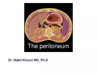
The peritoneum. General features The peritoneum is a thin serous membrane Consisting of: 1- Parietal peritoneum - lines the ant. Abdominal wall 2- Visceral peritoneum covers the viscera Peritoneum is continuous below with parietal peritoneum lining the pelvis 3- Peritoneal cavity

E N D
General features • The peritoneum is a thin serous membrane • Consisting of: 1- Parietal peritoneum -lines the ant. Abdominal wall 2- Visceral peritoneum • covers the viscera • Peritoneum is continuous below with parietal peritoneum lining the pelvis 3- Peritoneal cavity • the potential space between the parietal and visceral layer of peritoneum • in male, is a closed sac • but in the female, there is a communication with the exterior through the uterine tubes, the uterus, and the vagina ▪
Peritoneum……cont • Peritoneum cavity divided into Greater sac Lesser sac • Communication between them by the epiploic foramen
Lesser sac = omental bursa • Deep to lesser omentum • Behind the stomach • Between two layers of greater omentum • Under the diaphragm and liver • Deep to lesser opening (Epiploic opening)
Omental bursa……cont Walls: • Superior-peritoneum which covers the caudate lobe of liver and diaphragm • Anterior-lesser omentum, peritoneum of posterior wall of stomach, and anterior two layers of greater omentum
Omental bursa……cont • Inferior-conjunctive area of anterior and posterior two layers of greater omentum • Posterior-posterior two layers of greater omentum, transverse colon and transverse mesocolon, peritoneum covering posterior abdominal wall.
Omental bursa……cont • Left- spleen, gastrosplenic ligament splenorenal ligament • Right-omental foramen
Greater sac Deep to ant. Abdominal wall Below the diaphragm Above pelvic viscera Out to: Liver surround all the liver except bare area Stomach completely surrounded by peritoneum Transverscolon Greater omentum two layers of peritoneum from greater curvature of stomach Duodenum just the anterior surface covered by peritoneum Small intestine surrounds all the intestine & form mesentery
Greater sac Subdivided greater omentum into : Antero- superior part Postero - inferior part
Greater sac…..cont • Antero – superior divided by Falciformligament into: • Right part • Left part
Greater sac……cont Poster – inferior divided by mesentery & small intestine into: Right part Left part
Omental (epiploic)foramen • Position: • lies between the liver and duodenum • just above the first part of the duodenum • behind the lesser omentum • infront of the inferior vena cava • short, vertically flattened passage, about 3cm
Epiploic foramen…cont The omental bursa (lesser sac) communicates with the greater sac through the omental foramen.
Epiploic foramen Boundaries Anteriorlly -Free border of lesser omentum contain 1- Bile duct( Rt & ant) 2- Hepatic artery(Lt & anT) 3- Portal vein(post.) Posteriorly I.V.C Superiorly Caudate process of caudate lobe of liver Inferiorly First part of duodenum
Function of the peritoneum • Secretes a lubricating serous fluid that continuously moistens the associated organs • Fat storage • Defense role the presence of lymphatic vessels & nodes • Support viscera
Intraperitoneal viscera Interperitoneal viscera Retroperitoneal viscera The relationship between viscera and peritoneum • Intraperitoneal viscera • viscera is almost totally covered with visceral peritoneum • example, stomach, 1st & last inch of duodenum, jejunum, ileum, cecum, vermiform appendix, transverse and sigmoid colons, spleen and ovary
The relationship between viscera and peritoneum….cont Interperitoneal viscera Such organs are not completely wrapped by peritoneum one surface attached to the abdominal walls or other organs. Example liver, gallbladder, urinary bladder and uterus
The relationship between viscera and peritoneum Retroperitoneal viscera some organs lie on the posterior abdominal wall Behind the peritoneum they are partially covered by peritoneum on their anterior surfaces only Example kidney, suprarenal gland, pancreas, descending and ascending colon, upper 3rd of rectum duodenum, and ureter, aorta and I.V.C
The Peritoneal Reflections or folds Certain terms, often arbitrary, are commonly used for the peritoneal reflections. A peritoneal reflection that connects the intestine and body wall is usually named according to the part of the gut to which it is attached. For example, the reflection to jejunum and ileum is termed the mesentery, that to the transverse colon is the transverse mesocolon. Some peritoneal reflections between organs or between the body wall and organs, are termed ligaments or folds. Most of such ligaments or folds contain blood vessels. Broad peritoneal sheets associated with stomach are termed omenta.
1- Omenta: Two-layered fold of peritoneum that extends from stomach to adjacent organs Two omenta Lesser omentum Greater omentum
Lesser omentum • Two-layered fold of peritoneum - Extends from porta hepatis, fissure of ligamentum venosum and the diaphragm to lesser curvature of stomach and superior part of duodenum
Lesser omentum • Hepatogastric ligamentfrom porta hepatis to lesser curvature of stomach • Hepatoduodenal ligament • - Extends from porta hepatis • to superior part of • duodenum, • - at its free margine enclose 3 structures(3 key structures) • common bile duct Ant. • proper hepatic a At the Lt. of the common bile duct • hepatic portal v post.
Contents of lesser omentum Blood vessels Rt. & Lt. gastric vessels Lymph nodes & lymphatic vessels Fat Autonomic N.S sympathetic + parasympathetic (vagus nerve)
Greater omentum • It is the largest peritoneal fold. • It consists of a double sheet, folded on itself so that it is made up of four layers. • The anterior two layers descend from the greater curvature of stomach and superior part of duodenum and hangs down like an apron in front of coils of small intestine • then turn up on the back of itself, and ascend to the transverse colon . - the two layers are separated to cover the anterior and posterior surfaces of transverse colon. Then they form the transverse mesocolon
The upper part of the greater omentum which extends between the stomach and the transverse colon is termed the gastrocolic ligament. In adult, the four layers of greater omentum are frequently adhered together, and are found wrapped about the organs in the upper part of the abdomen
Contents of Greater omentum (between the descended layers) Gastroepiploic vessels Lymph nodes & lymphatic vessels Fat Autonomic N.S sympathetic + parasympathetic (vagus nerve)
Functions of greater omentum ① protective function: The greater omentum contains numerous fixed macrophages, which performs an important protective function. ② storehouse for fat: The greater omentum is usually thin, and presents a cribriform apperarance, but always contains some adipose tissue, which in fatty people is present in considerable quantity. ③ migration and limation: The greater omentum may limit spread of infection in the peritoneal cavity. Because it will migrate to the site of any inflammation in the peritoneal cavity and wrap itself around such a site, the greater omentum is commonly referred to as the “policeman” of the peritoneal cavity.
Lessor omentum Greater omentum
2- Mesenteries of the peritoneum -Two-layered fold of peritoneum that attach the intestines to the posterior abdominal wall
1- Mesentery of small intestine -suspends the small intestine from the posterior abdominal wall -Broad and a fan-shaped • Root of mesentery • 15 cm long • Directed obliquely from left side of L2 vertebra to right sacroiliac joint
Mesentery of small intestine….cont Contents of the mesentery • the jejunal and ileal branches of the superior mesenteric artery &veins • nerve plexuses • lymphatic vessels • the lymphatic nodes, • connective tissue • fat
2- Mesoappendix • Triangular mesentery-extends from terminal part of ileum to appendix • Appendicular artery runs in free margin of the mesoappendix
3. The transverse mesocolon It is a broad fold Connects the transverse colon to the anterior border of the pancreas. Contents - The blood vessels - Nerves - lymphatic's of the transverse colon.
4-Sigmoid mesocolon - It is a fold of peritoneum attaches the sigmoid colon to the pelvic wall. Contents The sigmoid vessels Lymphatic vessels Nerves The left Ureter descends into the pelvis behind its apex.
1. The ligaments of the liver ① The falciform ligament of liver ② The ligamentum teres hepatis ③ The coronary ligament ④ The right triangular ligament ⑤ The left triangular ligament ⑥ The hepatogastric ligament ⑦ The hepatoduonedenal ligament
Falciform ligament of liver • Consists of double peritoneal layer • Sickeleshape • Extends from anterior abdominal wall (umbilicus) to liver • Free border of the ligament contains Ligamentum teres(obliterated umbilical vein)
Coronary ligamentthe area between upper and lower layer of the coronary ligament is the bare area of liver which contract with the diaphragm; • Left and right triangular ligamentsformed by left and right extremity of coronary ligament
Hepatogastric ligament • Hepatoduodenal ligament
2- Ligaments of spleen • Gastrosplenic ligament - Connects the fundus of stomach to hilum of spleen. - Contents the short gastric & left gastroepiploic vessels pass through it. • Splenorenal ligament • extends between the hilum of spleen and left kidney. • Contents • The splenic vessel • Lymphatic vessels ,nodes & nerve • the tail of pancreas
Ligaments of spleen • Phrenicosplenic ligament • Splenocolic ligament
3- Ligaments of stomach • Hepatogastric ligament • Gastrosplenic ligament • Gastrophrenic ligament • Gastrocolic ligament • Gastropancrestic ligament
4. The suspensory ligament of duodenum Sometimes named Treitz ligament at the junction between duodenum & jejunum
5. The phrenicocolic ligament It is a fold of peritoneum which is continued from the left colic flexure to the diaphragm opposite the 10th and 12th ribs.
4- The Peritoneal Recesses & fossa In certain parts of the abdomen, peritoneal fold may bound recesses or fossae of the peritoneal cavity. At the junction between intraperitoneal and retro peritoneal organs These recesses are of surgical importance since they may become the site of internal herniae, that is, a piece of intestine may enter a recess and may be constricted (strangulated) by the peritoneal fold granding the entrance to the recess. From a surgical point of view the omental bursa can be considered to belong to this category, with its opening at the epiploic foramen, bounded in front by the free border of the lesser omentum. They are sometimes found in relation to the duodenum, cecum and sigmoid colon.
The Peritoneal Recesses & fossa ….cont 1. Duodenal Recesses The superior duodenal recess or fossa The inferior duodenal recess or fossa The paraduodenal recess or fossa The duodenojejunal recess or fossa 2. Cecal recesses The superior ileocecal or fossa The inferior ileocecal or fossa The retrocecal recesses or fossa The rectocolic recess or fossa 3. The intersigmoid recess