Download
1 / 92
1.15k likes | 2.18k Views
DENTAL CARIES. CONTENTS. INTRODUCTION EPIDEMIOLOGY DEFINITION CLASSIFICATION THEORIES OF CARIES CURRENT CONCEPTS IN CARIES ETIOLOGY HISTOPATHOLOGY CONCLUSION. INTRODUCTION. EPIDEMIOLOGY. Pre-historic man have decreased caries prevalence Decreased caries prevalence in

E N D
CONTENTS • INTRODUCTION • EPIDEMIOLOGY • DEFINITION • CLASSIFICATION • THEORIES OF CARIES • CURRENT CONCEPTS IN CARIES ETIOLOGY • HISTOPATHOLOGY • CONCLUSION
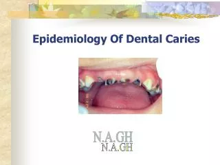
EPIDEMIOLOGY • Pre-historic man have decreased caries prevalence • Decreased caries prevalence in African and Asian communities • Factors affecting caries prevalence: • Race • Age • Sex • Familial
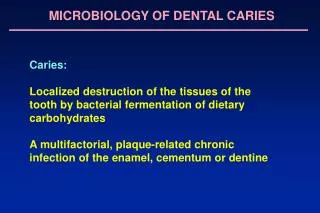
DEFINITION • “Dental caries is a microbial disease of the calcified tissues of the teeth, characterized by demineralization of the inorganic portion and destruction of the organic substance of the tooth” [Shaffer] • “Localized post eruptive pathologic process of external origin involving softening of the hard tissue and proceeding to the formation of a cavity” [WHO]
3. “An infectious microbiological disease of the teeth that results in localized dissolution and destruction of calcified tissues” [Sturdevant]
CLASSIFICATION • STURDEVANT Based on - Location - Extent - Rate of progression
According to location: a. Primary caries b. Caries of pit and fissure origin c. Caries of enamel smooth surface origin d. Backward caries e. Forward caries f. Residual caries g. Root surface caries h. Secondary (recurrent) caries
According to extent: a. Incipient (reversible) caries b. Cavitated (irreversible) caries • According to rate of progression: a. Acute (rampant) caries b. Chronic (slow or arrested) caries
G.V.Black’s Classification • Class-I: - caries on the occlusal surfaces of molars and premolars - occlusal 2/3 of the buccal and lingual surfaces of molars - lingual surfaces of the anterior teeth • Class-II: - lesions found on the proximal surfaces of molars and premolars • Class-III: - lesions found on the proximal surfaces of anterior teeth, but do not involve the incisal angle • Class-IV: - lesions found on the proximal surfaces of anterior teeth and involving incisal angle
Class-V: - lesions found on the gingival third of the facial and lingual surfaces of anterior and posterior teeth. • Class-VI: - were not included in Black’s classification - proposed by Siomon - lesions on the incisal edge and cusp tips of the teeth
GRAHAM MOUNT’S CLASSIFICATION Based on • Location of caries • Size of the carious lesion
PIT AND FISSURE CARIES • Limited to the – occlusal surfaces of molars and premolars - buccal pits of molars - lingual surfaces of maxillary anterior teeth • Poor self cleansing features • Usually occurs before smooth surface caries • Clinically - black or brown in color - slightly soft consistency - “catch” the tip of a fine explorer • Adjacent enamel appears bluish white • “Internal Caries”
Smooth Surface Caries • Develops on - proximal surfaces of the teeth - gingival third of the buccal and lingual surfaces (cervical caries) • Preceded by the formation of dental plaque. • Usually initiate just below the contact point. • Clinically- initially as faint white opacity or yellow brown pigmented area. • Adjacent enamel appears bluish white.
Cervical Caries • Appears as crescent shaped lesion. • May extend proximally. • Almost always an open cavity. • Lack of oral hygiene on the part of patient.
Backward Caries • Lateral spread of the lesion along the DEJ exceeds the caries in the contiguous enamel, caries extends into this enamel from the junction.
Forward Caries • Caries cone in enamel is larger or at least the same size as that in dentin
Residual Caries • Caries that remains in a completed cavity preparation • Not acceptable if - present at DEJ - prepared enamel wall
Root Surface Caries • In old age patients • Initiates at the surface of a mineralized dentin and cementum which have greater organic content • Usually have rapid clinical course
Recurrent (secondary) caries: • Occurs at the junction of the restoration and the cavosurface of the enamel • May extend beneath the restoration • Indicates unusual susceptibility to caries attack, poor cavity preparation, defective restoration. • Also indicates presence of microleakage.
Incipient (reversible) caries: • First evidence of caries activity in enamel • Clinically as white opaque region • Subsurface demineralization has occurred but no cavitation • May take up extrinsic stains • May undergo remineralization- called as “caries reversibility” or “consolidation” of early enamel carious lesion
Cavitated (irreversible) caries: • Lesion that has advanced into dentin with broken surface • Remineralization is not possible • Treatment include cavity preparation and restoring with suitable material.
Linear enamel caries (odontoclasia): • Atypical form of dental caries in primary dentition • Lesion predominates on the labial surface of the maxillary anterior teeth in the region of neonatal zone • Lesion is crescent shape • Increase caries susceptibility of posterior teeth.
Odontoclasia: - variant of linear enamel caries - results in gross destruction of the labial surfaces of incisor teeth - cause may be an inherent structural defect
Acute dental caries: • Rapid clinical course resulting in early pulp involvement • Frequently in children and young adults • Entry of lesion remains small while rapid spread along the DEJ • Clinically appears light yellow in colour • Pain is often present
Chronic dental caries • Slowly progressive lesion that involves pulp much later • Common in adults • Large entrance of the lesion • Dentin is stained deep brown • Moderate lateral spread of caries at DEJ • Pain is not a common clinical finding.
Rampant caries: • Sudden and rapid onset and almost uncontrollable destruction of teeth • Involves teeth that are ordinarily caries free (mandibular incisors) • Ten or more new increments of carious lesion in one year
Nursing Bottle (Infancy or Soother) Caries • Rapidly progressing caries affecting primary dentition usually during first 2 years of life • 4 maxillary anterior are affected first • If unchecked, maxillary and mandibular molars may also get involved • Lower anterior are spared (characteristic feature)
Adolescent caries: • Acute caries attack at 11-18 years of age • Lesion in teeth and surfaces that are relatively immune to caries • Small opening in enamel with extensive undermining • Rapid clinical course • Little or no secondary dentin formation
Arrested caries: • Caries which becomes static or stationary and does not show any tendency for progression • Almost exclusively occurs on occlusal surfaces • Both dentitions are affected • Lesion appears as large open cavity with lack of food retention • Superficially softened and decalcified dentin gets burnished and has brown stained polished appearance “Eburnation of dentin”
Xerostomia induced caries (radiation caries) • Complication of radiation therapy of oral cancer lesion • Radiation induced xerostomia produces caries conducive environment • Carious lesion develops as early as 3 months after onset of xerostomia • May be caused by other factors like salivary gland tumors, autoimmune diseases, prolong illness
Senile Caries • Caries activity that spurts up during the old age. • They are located exclusively on the root surfaces of the teeth. • Also seen in association with partial denture clasps. • Causes: gingival recession, decreased salivary secretion, poor oral hygiene.
Occult Caries / Hidden Caries • Not clinically diagnosed, but detected only on radiograph. • Seen in persons with low caries index suggestive of increased fluoride exposure. • Also called as fluoride bombs or fluoride syndrome
Simple caries: one surface is involved • Compound caries: two surfaces are involved • Complex caries: three or more surfaces are involved
ETIOLOGICAL THEORIES • Early theories • Endogenous theories • Exogenous theories
EARLY THEORIES • The Legend of the Worm: • According to an ancient Sumerian text, tooth ache was caused by a worm that drank the blood of the teeth and fed on the roots of the jaws • Antony Van Leeuwenhock (1700) the father of modern microscopy wrote a letter to the Royal Society of London describing little worms taken out of a corrupt tooth and said that they caused the pain in tooth ache.
ENDOGENOUS THEORY • Humoral Theory “dental caries is produced by internal action of acid and corroding humors”. • Four elemental fluids of the body-blood, phlegm, black bile and yellow bile • Vital Theory “tooth decay originated, like bone gangrene, from within the tooth itself”
EXOGENOUS THEORY • Chemical (Acid) theory: “ teeth are destroyed by acids formed in the oral cavity ” • Parasitic (Septic) theory: “ microorganisms are associated with carious process ”
Acidogenic Theory • Proposed by W.B.Miller 1890, most accepted. • “Acids formed due to the fermentation of dietary carbohydrates by oral bacteria leads to progressive decalcification of tooth structure with subsequent disintegration of organic matrix.”
He isolated micro-organisms from his experiments & stated that many were involved in the carious process. • 3 important factors which can influence process of tooth destruction in process of dental caries: Dietary carbohydrates, micro-organisms and acid
Proteolytic Theory • Proposed by Glottlieb in 1941. • “formation of dental caries is essentially proteolytic process. Bacteria present produce hydrolytic enzymes and cause proteolysis leading to the dissolution of organic substance.” • Microorganisms invade the organic substance first.
He did admit that, acid formation accompanied the proteolysis. • Yellow pigmentation of dental caries is because of pigment production by proteolytic organism. • Caries is initiated at slightly alkaline pH.
Proteolytic-chelation Theory • Proposed by Schwatz in 1955. • Stated that two processes are involved in caries – proteolysis & chelation.
Chelation is a process involving complexing of ions into a complexing substance by covalent bond which results in a highly stable, poorly dissociated and weekly ionized compound called chelate. • Bacterial attack on enamel is initiated by keratolytic microorganisms. • Eg: citrates, lactates • Chelates are always –ve.
Sucrose Chelation Theory • If there is a very high concentration of sucrose in mouth of a caries active individual, there can be formation of complexes like calcium saccharates, calcium complexing intermediaries, etc by action of phosphorylating enzymes • These complexes cause release of Ca, P ions from enamel and result in DC
Autoimmune Theory • Few odontoblast cells at specific sites within pulp of specific teeth are damaged by autoimmune mechanism • Due to this the defense capacity & integrity of of enamel and dentin in those specific areas are compromised and act as potential sites for caries development in future
Sulfatase Theory • Proposed by Pincus in 1951. • Bacterial sulfatases hydrolyses the mucoitin sulfate of enamel and chondroitin sulfate of dentin producing sulfuric acid that in turn causes decalcification. • Limitation: Sulfated polysacharide in enamel is very small and not readily accessible as a substrate for enzymatic degradation. So, this is highly unlikely hypothisis for the degragation of tooth enamel.
Levine’s theory • Proposed by Levine in 1977. • Established chemical relationship between enamel, plaque and factors which favors the movement of minerals between them. • Also called as “SEW – SAW” mechanism.
Said enamel demineralization and remineralization is a continuous process. • Movement of ions between enamel and plaque occurs in both direction which depend upon - plaque ph - calcium and phosphate ions at the interface - fluoride ion concentration
CURRENT CONCEPTS IN CARIES ETIOLOGY KEYE’S DIAGRAM VENN DIAGRAM