Download
1 / 39
460 likes | 1.03k Views
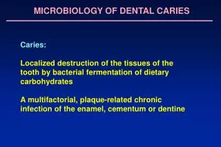
Dental Caries- Histoplathology. Dr. Rhythm Assistant Professor. CARIES OF DENTIN Begins with the natural spread of the process along the DEJ and rapid involvement of the dentinal tubules. The dentinal tubules act as tracts leading to the pulp (path for micro-organisms). Caries of Dentin.

E N D
Dental Caries- Histoplathology Dr. Rhythm Assistant Professor
CARIES OF DENTIN Begins with the natural spread of the process along the DEJ and rapid involvement of the dentinal tubules. The dentinal tubules act as tracts leading to the pulp (path for micro-organisms).
Early Dentinal Changes: -initial penetration of the dentin by caries dentinal sclerosis, -calcification of dentinal tubules and sealing off from further penetration by micro-organisms, -more prominent in slow chronic caries.
In the earliest stages, when only few tubules are involved, microorganisms may be found penetrating the tubules Pioneer Bacteria.
This initial decalcification involves the walls allowing them to distend as the tubules are packed with microorganisms. Each tubule is seen to be packed with pure forms of bacteria, eg., one tubule packed with coccal forms the other tubule with bacilli. 6
Advanced Dentinal Changes ; -decalcification of walls, confluence of the dentinal tubules, -tiny “liquefaction foci”, described by Miller are formed by the focal coalescing and breakdown of dentinal tubules. These are ovoid areas of destruction parallel to the course of the tubules which filled with necrotic debris and increase in size by expanding. The adjacent tubules are distorted and their course is bent due to this expansion. 7
As the microorganisms proceed further they are distanced from the carbohydrates substrate that was needed for the initiation of the caries. Thus the high protein content of dentin must favour the growth of the microorganisms. Therefore proteolytic organisms might appear to predominate in the deeper caries of dentin while acidophilic forms are more prominent in early caries. 9
Shape of the lesion is triangular with the apex towards the pulp and the base towards the enamel. Zone 1; Zone of Fatty Degeneration of Tome’s Fibers,(next to pulp) -due to degeneration of the odontoblastic process. This occurs before sclerotic dentin is formed and makes the tubules impermeable. 10
Zone 2; Zone of dentinal sclerosis, -deposition of Ca salts in the tubules. Zone 3; Zone of decalcification of dentin Zone 4; Zone of bacterial invasion Zone 5; Zone of decomposed dentin due to acids and enzymes. 11
Root Caries Root caries as defined by HAZEN, is a soft, progressive lesion that is found anywhere on the root surface that has lost its connective tissue attachment and is exposed to the environment. -the root surface must be exposed to the oral environment before caries can develop here. -Plaque and micro-organisms are essential for the cause and progression of the lesion, mostly Actinomyces, -micro-organisms invade the cementum either along the Sharpey’s fibers or between the bundles of fibers.
-spread laterally, since cementum is formed in concentric layers. -after decalcification of cementum, destruction of matrix occurs similar to dentin with ultimate softening and destruction of this tissue. -invasion of micro-organisms into the dentinal tunbules, finally leading to pulp involvement. -the rate is slower due to fewer dentinal tubules than crown area
Importance of Early Caries Detection Importance of early caries detection
Objectives of diagnosis: Identifying lesions requiring surgical treatment Identifying lesions requiring non-surgical treatment Persons at high risk for developing caries Thus diagnosis is done using: Clinical criteria Tools Newer refined diagnostic tools
VISUAL EXAMINATION Most widely used method, in dental offices, in clinical research and in epidemiological studies. • Quick, cheap and easy. • Should be performed on a dry, clean tooth, with good light, with a mirror. Wet Dry
TACTILE METHOD • Sturdevant’s (1985) : • Defects are best detected when an explorer placed into a pit or fissure provides tug-back or resistance to removal. • Subject of controversy: • Use of the explorer does not add anything to the detection yield of the examination. • The use of the explorer may at best be misleading and at worst be potentially damaging
RADIOGRAPHY The purpose of the radiograph is to detect lesions that are clinically hidden from careful visual examination. ADVANTAGES : • Discloses sites inaccessible to other methods • Detects at early , reversible stage • Depth of lesion can be evaluated and scored by index given by Grondahl et al (1977) • Permanent record
BITE WING RADIOGRAPHS When radiography is applied in the clinic for caries detection, the recommended technique is bitewing projection (Gröndahl, 1994). Disadvantages Not convincingly able to distinguish between cavitated and noncavitated surfaces (Nielsen et al., 1996)
XERORADIOGRAPHYBY CARLSON 1937 Image is recorded on an aluminium plate coated with a layer of selenium particles which have a uniform electrostatic charge X-rays cause selective discharge from the particles, forming a latent image, which is developed to a +ve image by developing Advantages: Twice as sensitive as conventional D-speed films “Edge enhancement”is possible
QLF, THE QUANTITATIVE LIGHT INDUCED FLUORESCENCE METHOD The science behind this phenomenon appears to be the increased fluorescence exhibited by cariogenic bacterial metabolites within the lesion, as well as the changed fluorescent nature of the lesion itself.
ENHANCED VISUAL TECHNIQUES FIBER OPTIC TRANS-ILLUMINATION( FOTI): Principle: decayed matter scatter light more strongly-lower index of light transmission Can be used in all surfaces, particularly useful at proximal lesions
Dyes can visualize a subject from its routine background by colour. The observation can be qualitative or quantitative. Dyes should be -safe for intra-oral use -stain the tissues that are diseased -should be easily removed DYES USED FOR CARIES DIAGNOSIS Vista red, Vistadental
DYES IN CARIES DETECTION Carious Enamel: Procion, Calcein, Brilliant Blue is used. • Dentin caries: Basic fuchsin , Methylene blue, Acid red. • Modified dye preparation uses iodine. • It measures enamel porosity in incipient carious lesions Bakhos et al.(1977) • Uses iodine as potassium iodide.
DIGITAL RADIOGRAPHY A digital imaging is an image formed and represented by a spatially distributed set of discrete sensors and pixels when viewed from a distance the image appear continuous, but on closer inspection it has individual pixels.
Caries detecting dyes stain infected carious dentin, but also stain the demineralized organic matrix of carious dentin, which should not be removed.
Q. 1 Bactria found in initial dentinal caries are called A. Initial bacteria B. Frontier bacteria C. Pioneer Bacteria D. Premier bacteria
Q.2 Dentinal sclerosis is seen in A. slowly progressing caries B. Acute caries c. Incipient caries D. irreversible caries
Q.3 Liquefaction foci can be seen in A. Advanced caries B. Initial caies C. Rampant caries D. Incipient caries
Q.4 Early caries is associated with A. Predominantly acidogenic bacteria B. Proteolytic bacteria C. Premier bacteria D. acidophillic bacteria
Q.5 Zone of fatty degeneration is A. Zone 1 B. Zone 2 C. Zone 3 D. zone 4
Q. 6 Root caries is caused due to A. Actinomyces B. Lactobacillus C. Streptococcus D. Staphylococcus
Q.7 QLF is based on a. Fluorescence b. Absorption c. Scatter d. Radiation
Q.8 Bite wing radiographs are best for a. Visualising perapical area b. Proximal caries c. Occlusal caries d. Root caries
Q.9 Root caries is caused by a. Actinomyces b. Lactobacilli c. Streptococcus d. Staphylococcus
Q.10 Dental floss is used for detecting a. proximal caries b. occlusal caries c. root caries d. enamel caries