Download
1 / 81
820 likes | 1.28k Views
Muscle. Topic 4-1 February 2014. Mid-term Exam. Friday, Feb. 14 th 9:00-9:50 am Mix multiple choice, short answer, fill in the blank, label a diagram Pathways are important. Lecture Overview. Muscle structure Sarcomeres Excitation-contraction coupling Ca2+ control of contraction

E N D
Muscle Topic 4-1 February 2014
Mid-term Exam • Friday, Feb. 14th • 9:00-9:50 am • Mix multiple choice, short answer, fill in the blank, label a diagram • Pathways are important
Lecture Overview • Muscle structure • Sarcomeres • Excitation-contraction coupling • Ca2+ control of contraction • Sliding filament model • Actino-myosin activity • Relaxation
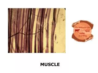
Muscle Cells • Two main types of muscle cells are based on the arrangement of actin and myosin • Striated (striped appearance) • Smooth (do not appear striped) Figure 5.16
endomysium perimysium Normal muscle structure and collagen distribution
endomysium perimysium
18 mo 27 mo 30 mo 33 mo 36 mo 39 mo Lushaj EB, Johnson JK, McKenzie D, Aiken JM. Sarcopenia accelerates at advanced ages in Fisher 344xBrown Norway rats. J Gerontol A Biol Sci Med Sci. 2008;63:921–7.
Striated Muscle Types Table 5.3
Myofibrils in Muscle Cells Figure 5.20
Myofibrils • Single, linear continuous stretch of interconnected sarcomeres (i.e., in series) • Have parallel arrangement in the cell • More myofibrils in parallel can generate more force
Striated Muscle Cell Structure • Thick and thin filaments arranged into sarcomeres • Repeated in parallel and in series • Side-by-side across myocyte • Causes striated appearance • End-to-end along myocyte
Sarcomeres • Structural features of sarcomeres • Z-disk • A-band (anisotropic band) • I-band (isotropic band) • M-line
Actin & Myosin • Major components of microfilaments • Used for movement and movement of cellular components (vesicles)
Actin • Monomers = G-actin • Polymers = F-actin • Can spontaneously assemble/disassemble
Actin polymerization Figure 5.9
Myosin • 17 different classes of myosin • Multiple isoforms within each class • Muscle myosin = type II • General organization • Head • Neck • Tail
Myosin Figure 5.12
Myosin Isoforms • Properties of isoforms affect contraction • Multiple isoforms of myosin II in muscle • Isoforms can change over time Table 5.4
Contractile Elements • Thick filaments • Polymers of myosin • ~300 myosin II hexamers • Thin filaments • Polymers of -actin • Ends capped by tropomodulin and CapZ to stabilize • Proteins troponin and tropomyosin on outer surface
Thick and Thin Filaments Figure 5.15
http://education.vetmed.vt.edu/curriculum/VM8054/Labs/Lab10/IMAGES/305-03%20SMALL.jpghttp://education.vetmed.vt.edu/curriculum/VM8054/Labs/Lab10/IMAGES/305-03%20SMALL.jpg
Three-Dimensional Structure of Sarcomere Figure 5.18
Sarcomeres Figure 5.17
Auxiliary Proteins • Nebulin • Along length of thin filament • Titin • Keeps thick filament centered in sarcomere • Attaches thick filament to Z-disk
Neurogenic Muscle Figure 5.25
http://scientopia.org/blogs/scicurious/files/2010/11/neuromuscular-junction.jpghttp://scientopia.org/blogs/scicurious/files/2010/11/neuromuscular-junction.jpg
http://fig.cox.miami.edu/~cmallery/150/neuro/neuromuscular-sml.jpghttp://fig.cox.miami.edu/~cmallery/150/neuro/neuromuscular-sml.jpg
Acetylcholine Figure 4.17
Acetylcholine • 1 of many NTs in autonomic system • Sole NT for motor division of the somatic system • 1st identified by Henry Dale http://t0.gstatic.com/images?q=tbn:ANd9GcR8mzV4vXbaOt8PSXQTlcSPmqJOOTMlz3TY2OO1_P2_F-K5QToy
Receptors for Acetylcholine • Cholinergic receptors • Nicotinic receptor • Ionotropic • Muscarinic receptor • Metabotropic • Linked to ion channel function via G-protein
Receptors for Acetylcholine Figure 4.29
Receptors for Acetylcholine Table 4.5
Depolarization • Neurogenic (“beginning in the nerve”) • Excited by neurotransmitters from motor nerves • Can have multiple (tonic) or single (twitch) innervation sites • Receptor is nicotinic • ionotropic
Action Potentials • APs along sarcolemma signal contraction • Na+ enters cell when Na+ channels open • Depolarization • Voltage-gated Ca2+ channel open • Increase in cytoplasmic [Ca2+] • Na+ channels close • K+ leaves cell when K+ channels open • Repolarization • Reestablishment of ion gradients by Na+/K+ ATPase and Ca2+ ATPase
Time Course of Depolarization Figure 5.24
T-Tubules and SR Figure 5.28
http://people.eku.edu/ritchisong/301images/Transverse_tubule.jpghttp://people.eku.edu/ritchisong/301images/Transverse_tubule.jpg
Ca2+ Channels and Transporters Figure 5.27