Download
1 / 44
440 likes | 568 Views
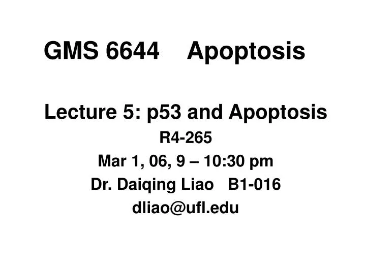
GMS 6644 Apoptosis. Lecture 5: p53 and Apoptosis R4-265 Mar 1, 06, 9 – 10:30 pm Dr. Daiqing Liao B1-016 dliao@ufl.edu. Tumor-suppressor genes.

E N D
GMS 6644 Apoptosis Lecture 5: p53 and Apoptosis R4-265 Mar 1, 06, 9 – 10:30 pm Dr. Daiqing Liao B1-016 dliao@ufl.edu
Tumor-suppressor genes Tumor-suppressor genes, function like brakes, keep cell numbers down, either by inhibiting progress through the cell cycle and thereby preventing cell birth, or by promoting programmed cell death (also called apoptosis). When cellular tumor suppressor genes are rendered non-functional through mutation, the cell becomes malignant. Examples are the gene encoding the retinoblastoma protein (Rb), inactivated in retinoblastomas, p53, and p16INK4a, which inhibits cyclin-dependent kinases and is inactivated in many different tumors.
Oncogenes Oncogenes stimulate appropriate cell growth under normal conditions, as required for the continued turnover and replenishment of the skin, gastrointestinal tract and blood, for example. Cells with mutant oncogenes continue to grow (or refuse to die) even when they are receiving no growth signals. Examples are Ras, activated in pancreatic and colon cancers, and Bcl-2, activated in lymphoid tumours. Amplification of oncogenes (more than their normal gene copy number) is also found in cancer: MDM2 is amplified in liposarcomas.
Roles of p53 in apoptosis p53 induces apoptosis through transcriptional activation of proapoptotic genes, such as Puma, Noxa, p53AIP1, Bax, Apaf-1 etc. It can also directly induce apoptosis by localizing to mitochondria via interaction with Bcl-2 family protein Bcl-xL and facilitating Bax oligomerization Reading: Vousden and Lu: Nature Reviews Cancer, 2002, 2:594-604.
p53 and of apoptosis Ref: Mol. Cell, 2003, 11(3):552-4
p53 and apoptosis Ref: Cell, 2002, 108:153-164
Tumor Suppressor p53 • First identified as a protein associated with viral oncogenes • Mutated/inactivated in a majority of human cancers • Integrates numerous signals that control cell life and death • A common denominator in human cancer • Understanding functions and regulation of p53 is of great importance in cancer biology and cancer therapy
p53 is a sequence-specific DNA binding protein p53 central core-domain interacts directly with DNA p53 binding sites consist of four copies of the pentamer consensus sequence PuPuPuC(A/T). The pentamers are oriented in alternating directions. A short stretch of sequence up to 13 bp may be inserted between the pairs of pentamers. The p53 target genes in the human genome usually carry the consensus sequence. 3. Amino acid residues in the core-domain that are critical for DNA-binding are among the “hot-spots” of tumor-derived p53 mutations, attesting to the importance of DNA-binding for p53’s tumor suppression function.
Structure of p53 core-domain Ref: Science, 265:346-355, 1994
Structure of the p53 OD Science 1995, 67:1498-1502.
The p53-MDM2 feedback loop MDM2 binds to p53 N-terminal transactivation domain and inhibits p53-dependent transcription. 2. MDM2 is a transcription target of p53. MDM2 is an E3 ubiquitin ligase of p53, thus targeting p53 for proteolytic degradation. 4. MDM2 knockout is lethal for mouse embryonic development, but simultaneous deletion of p53 and MDM2 genes rescues MDM2-KO, thus confirming the in vivo genetic interaction of these two proteins.
Structure of MDM2-p53 complex Science 1996 274:948-953
p53 and apoptosis p53AIP1 Apaf-1
p53 in apoptosis p53 mediates apoptosis in response to DNA damage, oncogene expression (adenovirus E1A, myc etc.), or withdrawal of growth factors 2. Overexpression wild-type of p53 leads to apoptosis 3. p53 can induce the expression of proapoptotic genes, such as Bax (ref Cell, 80:293) and p53AIP1 (ref: Cell, 102:849)
p53 in apoptosis 4. p53 can also repress transcription of certain genes, and it has been proposed that the repression function may also be required for apoptosis (ref: Genes Dev 13:2490-501) 5. In vivo, p53 transactivation mutant is defective in inducing apoptosis, at least for some cell types (ref: EMBO J. 19:4967-4975)
Physiological relevance of p53-induced apoptosis • Suppress oncogene-induced transformation • Inhibit tumor growth and progression • Remove cells with severe DNA damage • Effectiveness of cancer chemotheraphy correlates with the ability to induce p53-dependent apoptotic response
Tumor-derived mutations affecting apoptosis Ref: Cell, 2002, 108:153-164
Tumor-derived mutations affecting apoptosis Ref: Cell, 2002, 108:153-164
Tumor-derived mutations affecting apoptosis Ref: Cell, 2002, 108:153-164
Tumor-derived mutations affecting apoptosis Ref: Cell, 2002, 108:153-164
Tumor-derived mutations affecting apoptosis Ref: Cell, 2002, 108:153-164
Tumor-derived mutations affecting apoptosis Ref: Cell, 2002, 108:153-164
p53 directly mediates mitochondrial mechanism of apoptosis Early studies indicate that transactivation-defective mutants of p53 are capable of inducing apoptosis, implying a transcription-independent role for p53 in apoptosis. DNA-damage leads to mitochondrial translocation of p53. p53 binds to Bcl-2 family protein Bcl-xL to influence cytochrome c release. p53 directly activates the proapoptotic Bcl-2 protein Bax in the absence of other proteins to permeabilize mitochondria and engage the apoptotic program. p53 can release both proapoptotic multidomain proteins and BH3-only proteins [Proapoptotic Bcl-2 family proteins that share only the third Bcl-2 homology domain (BH3)] that are sequestered by Bcl-xL. Ref: Mol. Cell, 2003, 11:577-90; Science, 2004, 303:1010-4.
p53 is found in mitochondria following DNA damage Ref: Mol. Cell, 2003, 11:577-90
Recombinant p53 causes rapid cytochrome c release from mitochondria Ref: Mol. Cell, 2003, 11:577-90
Cooperation of endogenous cytoplasmic p53 with Bax to induce apoptosis in the absence of p53 nuclear activity Ref: Science, 2004, 303:1010-4.
How p53 functions in mitochondria to induce cell death 1. p53 or Bax alone does not permeabilize membrane, but they together can do so. 2. p53 facilitates Bax oligomerization. 3. p53 binds to Bcl-xL, but not to Bax. 4. p53-Bcl-xL interaction releases Bax. 5. Released Bax forms oligomers in mitochondrial membrane, leading cytochrome c release and apoptosis. 6. The proline-rich domain (aa 62-91 in mouse) of p53 is required for this effect. Ref: Science, 2004, 303:1010-4.
p53 binds to Bcl-xL and releases Bax Bax is sequestered by Bcl-xL and inactive Bax is released by p53 from Bcl-xL and forms oligomers, leading to apoptosis Ref: Science, 2004, 303:1010-4.
p53 and Puma in apoptosis Ref: K. Vousden, Science, 2005
Viral oncogenes and the p53 network Ref: Nature408, 307 - 310 (2000)
Viral oncogenes inhibit p53 functions 1. p53 was first identified as binding protein of SV40 large T antigen in 1979, which interacts with p53 core domain 2. Adenovirus oncoprotein E1B 55-kDa binds to p53 and inhibits p53 transactivation activity 3. HPV E6 binds to p53 and targets it for ubiquitin-mediated degradation.
Ad12 E1B and p53 colocalize in the cytoplasmic bodies Ref: Zhao&Liao. J. Virol., 2003,77:13171-13181.
Inhibition of p53-mediated apoptosis by E1B 55-kDa through cytoplasmic sequestration • Ad12 E1B 55-kDa forms large and filamentous cytoplasmic body • p53 localizes to the E1B cytoplasmic body, and its N-terminal TAD is required for this localization • Sequestration of p53 in the cytoplasmic body correlates with the ability of E1B 55-kDa to repress p53-dependent apoptosis
Using p53 to kill cancer cells The p53 protein is a tumor suppressor — it keeps cell numbers down by stopping cells from multiplying or by promoting cell death. Loss of p53 occurs in most human cancers, so it would be useful to be able to restore its function. Several innovative strategies have been suggested: • Introduce normal p53 genes into a cancer cell with mutant p53. • Introduce a small compound that converts mutant p53 proteins from an abnormal to a normal shape. • Add a protein that attaches itself to mutant p53 and kills cells. • Stimulate the host's immune response to mutant p53 peptides.
Using p53 to kill cancer cells • Introduce drugs that disrupt the interaction between the MDM2 or E6 proteins and p53. (MDM2 and E6 negatively regulate p53; they are present at abnormally high levels in some cancer cells, so 'quench' any normal p53.) • Introduce the adeno-associated virus, which mimics damaged DNA. Cells with mutant p53 cannot activate the usual p53-dependent 'checkpoint' that is induced by DNA damage, and eventually die. • Infect cells with viruses that can replicate only in cells without normal p53; the viruses kill these cells.
AAV kills cancer cells Ref: Nature, 412, 865 - 866 (2001)
p53 in DNA repair and apoptosis Ref: Bensaad & Vousden, Nature Med, 2005 ROS: reactive oxygen species
Summary p53 is a tumor-suppressor protein that induces apoptotic cell death in response to oncogenic stress. Malignant progression is dependent on loss of p53 function, either through mutation in the TP53 gene (which encodes p53) itself or by defects in the signaling pathways that are upstream or downstream of p53. Mutations in TP53 occur in about half of all human cancers, almost always resulting in the expression of a mutant p53 protein that has acquired transforming activity. p53-induced apoptosis depends on the ability of p53 to activate gene expression. p53 can also directly trigger the apoptotic response, by interacting with Bcl-2 family protein. The apoptotic and cell-cycle arrest activities of p53 can be separated, and apoptotic cofactors that play a specific role in allowing p53-induced death are being identified. Ref: Nature Reviews Cancer, 2, 594 - 604 (2002)
Summary Phosphorylation of p53 regulates its ability to activate the expression of apoptotic target genes, and other post-translational modifications such as acetylation might also have a role. In tumors that retain wild-type p53, the apoptotic response might be hindered by defects in the apoptotic cofactors. These, therefore, represent additional targets for the design of therapeutics that are aimed at reactivating p53-mediated apoptosis in cancer cells. Ref: Nature Reviews Cancer, 2, 594 - 604 (2002)