Download
1 / 45
460 likes | 532 Views
Gene Expression and Development. Final Exam. Sunday, May 27, 8:30-11:30 a.m. Here – SMC A110 Some review during class on Friday. Important Readings for Gene Expression and Development. Campbell chapter 18.4 Campbell chapter 21.6 Matt Ridley, Genome , chapter 12 ‘Self-Assembly’. Overview.

E N D
Final Exam • Sunday, May 27, 8:30-11:30 a.m. • Here – SMC A110 • Some review during class on Friday
Important Readings for Gene Expression and Development • Campbell chapter 18.4 • Campbell chapter 21.6 • Matt Ridley, Genome, chapter 12 ‘Self-Assembly’
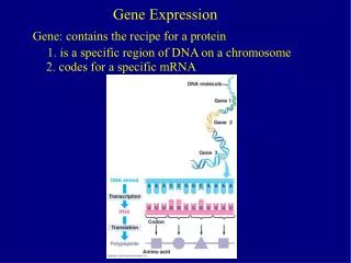
Overview • Both prokaryotes and eukaryotes alter their patterns of gene expression in response to changes in environmental conditions. • Multicellular eukaryotes also develop and maintain multiple cell types. • Each cell type contains the same genome but expresses a different subset of genes. • During development, gene expression must be carefully regulated to ensure that the right genes are expressed only at the correct time and in the correct place.
Fig. 18-14 How do we go from a fertilized egg to a fully developed individual? (a) Fertilized eggs of a frog (b) Newly hatched tadpole
(a) Animal development. Most animals go through some variation of the blastula and gastrula stages. The blastula is a sphere of cells surrounding a fluid-filled cavity. The gastrula forms when a region of the blastula folds inward, creating a tube—a rudimentary gut. Once the animal is mature, differentiation occurs in only a limited way—for the replacement of damaged or lost cells. Gut Cell movement Zygote (fertilized egg) Eight cells Blastula (cross section) Gastrula (cross section) Adult animal (sea star) Cell division Morphogenesis Observable cell differentiation (b) Plant development. In plants with seeds, a complete embryo develops within the seed. Morphogenesis, which involves cell division and cell wall expansion rather than cell or tissue movement, occurs throughout the plant’s lifetime. Apical meristems (purple) continuously arise and develop into the various plant organs as the plant grows to an indeterminate size. Seed leaves Shoot apical meristem Root apical meristem Zygote (fertilized egg) Two cells Embryo inside seed Plant Some key stages of development in animals and plants
The mantra for development: • Division • Morphogenesis • Differentiation Rather like – replication, transcription, translation for Mo Bio
Differential gene expression leads to different cell types and multicellularity • In the development of most multicellular organisms, a single-celled zygote gives rise to cells of many different types. • Each type has a different structure and corresponding function. • Cells of different types are organized into tissues, tissues into organs, organs into organ systems, and organ systems into the whole organism. • Thus, the process of embryonic development must give rise not only to cells of different types but also to higher-level structures arranged in a particular way in three dimensions.
The genetic program and development • As a zygote develops into an adult organism, its transformation results from three interrelated processes: cell division, cell differentiation, and morphogenesis. • Through a succession of mitotic cell divisions, the zygote gives rise to many cells. • Cell division alone would produce only a great ball of identical cells. • During development, cells become specialized in structure and function, undergoing cell differentiation. • Different kinds of cells are organized into tissues and organs. • The physical processes that give an organism its shape constitute morphogenesis, the “creation of form.” • Cell division, morphogenesis, and cell differentiation have their basis in cellular behavior.
How similar genetically are the brown rat and the house mouse?
The brown rat and the house mouse are identical at 67% of their euchromatic DNA
Chimpanzees and humans are genetically identical at 94% of their DNA
Fig. 18-14 How do we go from a fertilized egg to a fully developed individual? (a) Fertilized eggs of a frog (b) Newly hatched tadpole Division, Morphogenesis, and Differentiation
Early development of the zygote • Different sets of activators present in different cell types • One important source of information early in development is the egg’s cytoplasm, which contains both RNA and proteins encoded by the mother’s DNA, distributed unevenly in the unfertilized egg. • Maternal substances that influence the course of early development are called cytoplasmic determinants.
Cytoplasmic determinants • These substances regulate the expression of genes that affect the developmental fate of the cell. • After fertilization, the cell nuclei resulting from mitotic division of the zygote are exposed to different cytoplasmic environments. • The set of cytoplasmic determinants a particular cell receives helps determine its developmental fate by regulating expression of the cell’s genes during cell differentiation.
Fig. 18-15a Unfertilized egg cell Sperm Nucleus Fertilization Two different cytoplasmic determinants Zygote Mitotic cell division Two-celled embryo (a) Cytoplasmic determinants in the egg
More early development of the zygote • The other important source of developmental information is the environment around the cell, especially signals impinging on an embryonic cell from nearby cells. • In animals, these signals include contact with cell-surface molecules on neighboring cells and the binding of growth factors secreted by neighboring cells. • These signals cause changes in the target cells, a process called induction. • The molecules conveying these signals within the target cells are cell-surface receptors and other proteins expressed by the embryo’s own genes. • The signal molecules send a cell down a specific developmental path by causing a change in its gene expression that eventually results in observable cellular changes.
Fig. 18-15b NUCLEUS Early embryo (32 cells) Signal transduction pathway Signal receptor Signal molecule (inducer) (b) Induction by nearby cells
Cell Differentiation • During embryonic development, cells become visibly different in structure and function as they differentiate. • The earliest changes that set a cell on a path to specialization show up only at the molecular level. • Molecular changes in the embryo drive the process, called determination, which leads to the observable differentiation of a cell. • Once it has undergone determination, an embryonic cell is irreversibly committed to its final fate. • If a determined cell is experimentally placed in another location in the embryo, it will differentiate as if it were in its original position. • The outcome of determination—observable cell differentiation—is caused by the expression of genes that encode tissue-specific proteins. • These proteins give a cell its characteristic structure and function.
Cell Differentiation cont’d • Cells produce the proteins that allow them to carry out their specialized roles in the organism. • For example, liver cells specialize in making albumin, while lens cells specialize in making crystalline. • Skeletal muscle cells have high concentrations of proteins specific to muscle tissues, such as a muscle-specific version of the contractile proteins myosin and actin, as well as membrane receptor proteins that detect signals from nerve cells. • Muscle cells develop from embryonic precursors that have the potential to develop into a number of alternative cell types. • Although the committed cells are unchanged, they are now myoblasts. • Eventually, myoblasts begin to synthesize muscle-specific proteins and fuse to form mature, elongated, multinucleate skeletal muscle cells.
Fig. 18-16-1 Nucleus Master regulatory gene myoD Other muscle-specific genes DNA Embryonic precursor cell OFF OFF
Fig. 18-16-2 Nucleus Master regulatory gene myoD Other muscle-specific genes DNA Embryonic precursor cell OFF OFF OFF mRNA MyoD protein (transcription factor) Myoblast (determined)
Fig. 18-16-3 Nucleus Master regulatory gene myoD Other muscle-specific genes DNA Embryonic precursor cell OFF OFF OFF mRNA MyoD protein (transcription factor) Myoblast (determined) mRNA mRNA mRNA mRNA Myosin, other muscle proteins, and cell cycle– blocking proteins MyoD Another transcription factor Part of a muscle fiber (fully differentiated cell)
Pattern formation and the embryo body plan • Cytoplasmic determinants and inductive signals contribute to pattern formation, the development of spatial organization in which the tissues and organs of an organism are all in their characteristic places. • Pattern formation begins in the early embryo, when the major axes of animals and plants are established. • Before specialized tissues and organs form, the relative positions of a bilaterally symmetrical animal’s three major body axes (anterior-posterior, dorsal-ventral, right-left) are established. • The molecular cues that control pattern formation, positional information, are provided by cytoplasmic determinants and inductive signals. • These signals tell a cell its location relative to the body axes and to neighboring cells and determine how the cell and its progeny will respond to future molecular signals.
Pattern formation in Drosophila • Studies of pattern formation in Drosophila melanogaster have established that genes control development and have identified the key roles of specific molecules in defining position and directing differentiation. • Combining anatomical, genetic, and biochemical approaches in the study of Drosophila development, researchers have discovered developmental principles common to many other species, including humans. • Fruit flies and other arthropods have a modular construction. • An ordered series of segments make up the three major body parts: the head, thorax (with wings and legs), and abdomen. • Cytoplasmic determinants in the unfertilized egg provide positional information for two developmental axes (anterior-posterior and dorsal-ventral axis) before fertilization.
Fig. 18-17a Thorax Head Abdomen 0.5 mm Dorsal Right BODY AXES Posterior Anterior Left Ventral (a) Adult
Fig. 18-17b Follicle cell Egg cell developing within ovarian follicle 1 Nucleus Egg cell Nurse cell Egg shell Unfertilized egg 2 Depleted nurse cells Fertilization Laying of egg Fertilized egg 3 Embryonic development Segmented embryo 4 0.1 mm Body segments Hatching Larval stage 5 (b) Development from egg to larva
Pattern formation in Drosophila • Nüsslein-Volhard and Wieschaus studied segment formation in the late 1970s • They created mutants, conducted breeding experiments, and looked for corresponding genes • Breeding experiments were complicated by embryonic lethals, embryos with lethal mutations • They found 120 genes essential for normal segmentation
wildtype Wieschaus and Nüsslein-Volhard looked for mutants that affect the fly body plan
Axis Establishment • Maternal effect genes encode for cytoplasmic determinants that initially establish the axes of the body of Drosophila • These maternal effect genes are also called egg-polarity genes because they control orientation of the egg and consequently the fly
Fig. 18-17b Follicle cell Egg cell developing within ovarian follicle 1 Nucleus Egg cell Nurse cell Egg shell Unfertilized egg 2 Depleted nurse cells Fertilization Laying of egg Fertilized egg 3 Embryonic development Segmented embryo 4 0.1 mm Body segments Hatching Larval stage 5 (b) Development from egg to larva
Bicoid: A Morphogen Determining Head Structures • One maternal effect gene, the bicoid gene, affects the front half of the body • An embryo whose mother has a mutant bicoid gene lacks the front half of its body and has duplicate posterior structures at both ends
Figure 18.21 Head Tail A8 T2 T1 A7 T3 A6 A5 A1 A4 A2 A3 Wild-type larva 250 m Tail Tail A8 A8 A7 A6 A7 Mutant larva (bicoid)
Fig. 18-19b RESULTS Fertilization, translation of bicoid mRNA Anterior end 100 µm Bicoid mRNA in mature unfertilized egg Bicoid protein in early embryo
Fig. 18-19b RESULTS Fertilization, translation of bicoid mRNA Anterior end 100 µm Bicoid mRNA in mature unfertilized egg Bicoid protein in early embryo
Figure 18.22 100 m RESULTS Anterior end Fertilization,translation ofbicoid mRNA Bicoid mRNA in matureunfertilized egg Bicoid protein inearly embryo Bicoid mRNA in matureunfertilized egg Bicoid protein inearly embryo
Fig. 18-19c CONCLUSION Nurse cells Egg bicoid mRNA Bicoid mRNA in mature unfertilized egg Developing egg Bicoid protein in early embryo
This phenotype suggests that the product of the mother’s bicoid gene is concentrated at the future anterior end • This hypothesis is an example of the gradient hypothesis, in which gradients of substances called morphogens establish an embryo’s axes and other features
The bicoid research is important for three reasons: – It identified a specific protein required for some early steps in pattern formation – It increased understanding of the mother’s role in embryo development – It demonstrated a key developmental principle that a gradient of molecules can determine polarity and position in the embryo
Genetic Analysis of Early Development • Edward B. Lewis, Christiane Nüsslein-Volhard, and Eric Wieschaus won a Nobel Prize in 1995 for decoding pattern formation in Drosophila • In the 1940s Lewis discovered the homeotic genes, which control pattern formation in late embryo, larva, and adult stages
Homeotic Genes • Lewis was able to demonstrate that bizarre developmental mutations could be mapped on to the Drosophila chromosome map, providing the first concrete evidence that genes somehow direct the developmental process
Figure 18.20 Eye Leg Antenna Mutant Wild type