Download
1 / 17
180 likes | 310 Views
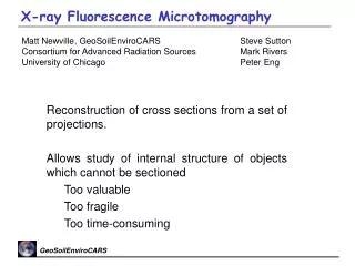
The Characterization of Uncertainties and Artifacts in X-Ray Microtomography. Tony Evershed Dental Biophysics Group, Institute of Dentistry. What is XMT?. Tomography, from Greek tomos (‘section’) and graphos (‘to write’). 2-3D representation based on a large number of projections.

E N D
The Characterization of Uncertainties and Artifacts in X-Ray Microtomography • Tony Evershed • Dental Biophysics Group, Institute of Dentistry
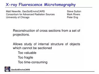
What is XMT? • Tomography, from Greek tomos (‘section’) and graphos (‘to write’). • 2-3D representation based on a large number of projections. • Tens-of-microns spatial resolution. • Attenuation coefficient resolution sufficient for mineral-content analysis.
XMT at QMUL • MuCAT Systems 1 and 2. • Cone-beam XMT with time-delay integrating detectors. • Based on COTS infrastructure with in-house software and detector hardware.
Cone Beam Tomography Image: Wikipedia (released into Public Domain)
Cone Beam Tomography Image: Wikipedia (released into Public Domain)
QMT at QMUL - TDI • Means of averaging pixel sensitivity. • Charge-coupled devices move charge in ‘steps’ by switching voltage at each pixel. • Synchronization of step frequency to sample movement. Animation: Michael Schmidt (released under GFDL.)
Applications of XMT Video: Dr G R Davis Examining decayed or damaged scrolls.
Image: F Ahmed Applications of XMT Analysis of biomaterial and artificial structures.
Applications of XMT Video: Dr G R Davis Mineralization studies in hard tissue.
Sources of Artifacts • Geometrical artifacts • Centre-of-rotation errors. • Specimen motion errors. • Focus: grayscale artifacts. • Beam Hardening • Scattering.
Artifacts: Beam Hardening • Arises from use of polychromatic radiation. • Materials do not follow Beer’s law: I = I0 e-μx. • Materials absorb ‘soft’ X-rays preferentially. • Beam becomes ‘harder’ and more penetrating. Image: Dr G R Davis
Observed LAC Artifacts: Beam Hardening • ‘Cupping’ artifact from beam hardening.
Artifacts: Beam Hardening • Correction: compare ideal Beer-law case with a known material added to the sample. • Multi-mode samples complicate matters. Image: Dr G R Davis
Artifacts: Scatter • Instead of being absorbed, photons may be deflected. • Compton (incoherent) scattering, from outer electron shells, largely responsible. • Increases level of noise in the reconstruction, particularly near high-attenuation regions. • Decreases contrast ratio in the reconstruction.
Artifacts: Scatter • Correction: none at present at QMUL. • Beam-hardening correction also corrects for some scatter. • Level of scatter can be determined from projection borders (outside cone beam.) • Monte Carlo modelling of virtual phantoms using Geant4 transport code.
Conclusion: Research Outcomes for QMUL • Reconstruction developments fed into existing MuCAT systems. • MuCAT 3 next-generation scanner. • Improved spatial resolution. • Larger sample capacity. • In tender, for delivery during 2012.
Acknowledgements • Supervisors: • Dr Graham Davis (Institute of Dentistry) • Dr Andrea Cavallaro (School of Electronic Engineering and Computer Science). • Post-doc: • Dr David Mills. • Engineering and Physical Sciences Research Council grant EP/G007845/1.