Download
1 / 32
391 likes | 472 Views
Pathophysiology of Cardiovascular System. Lecture # 3. Mrs.Iman Alajeyan ialajeyan@ksu.edu.sa. Circulation of blood. The human circulatory system is a two-part system :

E N D
Pathophysiology ofCardiovascular System Lecture # 3 Mrs.ImanAlajeyanialajeyan@ksu.edu.sa
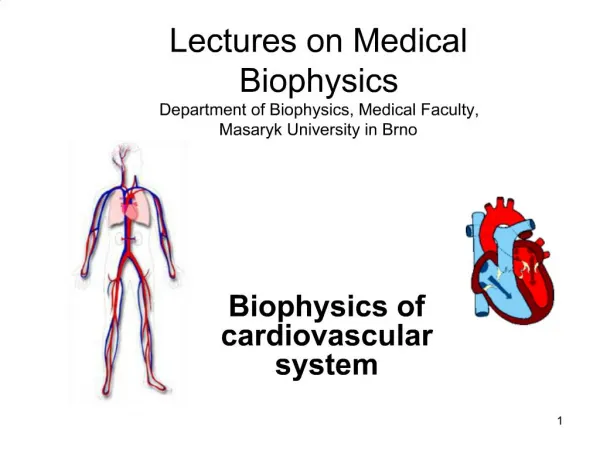
Circulation of blood The human circulatory system is a two-part system : The systemic circulation, the blood circulates into the body’s systems, bringing oxygen to all its organs, structures and tissues and collecting carbon dioxide waste. (between the heart and all other body tissues) The pulmonary circulation, the blood circulates to and from the lungs, to release the carbon dioxide and pick up new oxygen. (between the heart and the lungs) The systemic cycle is controlled by the left side of the heart, the pulmonary cycle by the right side of the heart.
Circulatory System It is a system of closed tubes through which blood circulates. It is formed of the heart and the blood vessels. Responsible for transporting oxygen, nutrients, hormones, and cellular waste products throughout the body Parts of the Circulatory System The circulatory System is divided into three major parts: 1.The Heart 2.The Blood 3.The Blood Vessels
Functions of different parts of the cardiovascular system: 1◘ The heart It is a muscle about the size of your fist. The heart is located in the center of your chest slightly to the left. Acts as a pump which provides the driving force for the circulation.
2◘ Blood Vessels Blood vessels are the body’s highways that allow blood to flow quickly and efficiently from the heart to every region of the body and back again There are three major types of blood vessels: arteries, capillaries and veins Arteries and Arterioles Arteries are blood vessels that carry blood away from the heart. Blood carried by arteries is usually highly oxygenated Arterioles are narrower arteries that branch off from the ends of arteries and carry blood to capillaries.
Capillaries: Are tiny blood vessels. Capillaries connect arteries to veins. Nutrients, oxygen and wastes pass in and out of your blood through the capillary walls. Venules and Veins: • Veins are blood vessels which carry blood to the heart. • Venules are small blood vessels which collect blood from capillaries. They progressively join together to form larger veins.
The heart The heart is composed of two halves (right and left), each half is formed of two chambers, an atrium and a ventricle. The atria Are relatively thin walled in relation to the ventricles. They receive and conduct passively the blood from the big veins to the ventricles. The ventricles The left ventricle is much thicker than the right ventricle. The main function of the ventricle is to pump blood into the circulatory system.
◘ Valves of the heart: The valves of the heart direct the flow of blood through the heart and into the large arteries and prevent backflow. A) Atrio-ventricular (A-V) valves: (mitral and tricuspid) These valves are present between atria and ventricles B) Semilunar valves: (aortic and pulmonary) These valves guard the opening between the ventricles and the large arteries, the aorta and the pulmonary arteries.
II. Pacemaking and conducting system: These is a group of specialized cardiac muscle cells in the walls of the heart which are specialized for rhythmic generation and conduction of cardiac impulses. This system consists of the following structures: (1) Sino-atrial (S.A) node, Is present in the right atrial wall at the junction of the superior vena cava with the right atrium. S.A node normally initiates the heart beats, so it is called the pacemaker of the heart.
2) inter-nodal tracts of the heart: • Three inter-nodal tracts, which conduct impulses from the S-A node to the A-V node.
(3) The atrio-ventricular (A-V) node, Is present on the right side of the inter-atrial septum at the atrio ventricular junction. (4) The bundle of His,) AV bundle) Originates from the A-V node then passes to the upper border of the inter-ventricular septum, where it divides into right and left bundle branches.
5) The bundle branches, Right and left branches, • The bundle branches spread down toward the apex of its ventricle where they are reflected upwards along the lateral walls of the ventricle. (6) The Purkinje fibers; The bundle branches subdivide into a network of rapidly conducting fibers called the Purkinje fibers.
The heartbeat is created by an electrical signal that starts in the right atrium. The signal is produced in the sinus node. The electrical signal moves down through the heart to the atrioventricular (AV) node. From the AV node, the electrical current travels to the ventricles along special fibers embedded in the heart walls ( bundle branch then to the Purkinje fibers). When the current arrives in the ventricles, they contract and pump blood out to the body https://www.youtube.com/watch?v=II5RPs1hlGI https://www.youtube.com/watch?v=RYZ4daFwMa8
Cardiac Output (COP) Cardiac Output (COP); is the total volume of blood pumped by each ventricle per minute. It equals 5-6 liters /min in adult during rest. COP of left side equals COP of right side. Stroke volume (S.V); is the volume of blood pumped by each ventricle per beat. COP = SV X HR. . Heart rate is the number of times your heart beats per minute.
Arterial blood pressure (ABP) Arterial blood pressureis the lateral pressure exerted by the blood on the arterial wall. During each cardiac cycle the ABP oscillates between systolic (maximum) and diastolic (minimum) pressure. Systolic blood pressureThe blood pressure when the heart is contracting. It is specifically the maximum arterial pressure during contraction of the left ventricle of the heart. Normally it is 120 mm Hg and ranges from 100 – 140 mm Hg). Diastolic blood pressureis specifically the minimum arterial pressure during relaxation and dilatation of the ventricles of the heart when the ventricles fill with blood. Normally it is 80 mm Hg and ranges from 60-90 mm Hg.
Rheumatic fever: • Acute recurrent or chronic inflammation of the heart that may affect endocardium, myocardium and cardiac valves • May occur following throat infection with group A beta-hemolytic streptococci bacteria • It is a disease of school-age children (5- to 15-year-old ) Symptoms Symptoms usually appear two to four weeks after your child has been diagnosed with strep throat. Common symptoms of strep throat include:
.•Fever •Painful and tender joints — most often the ankles, knees, elbows or wrists; •Pain in one joint that migrates to another joint •Red, hot or swollen joints •Small, painless nodules beneath the skin •Chest pain •Fatigue •Heart murmur ( unusual sound heard during a heartbeat.)
Disorders of cardiac valves Causes: 1-Inflammation:e.g, rheumatic fever 2-Damage: e.g, ischemia or direct trauma 3- Congenital Disorders in cardiac valves may lead to 2 types of changes : stenosis (is an abnormal narrowing) and incompetence (the valve does not close properly)
Cardiac arrhythmias Normal cardiac impulse conduction: occurs through: 1- Sinoatrial (SA) node: pace maker of heart 2- Atrioventricular (AV) node 3- Atrioventricular bundle (bundle of Hiss) 4-Left and right bundle branches 5-Purkinje fibers Heart rate • Normal heart rate is about 100-60 beats/min. and is regular • Heart rate below 60 beats/min is known as bradycardia. • Heart rate above 100 beats/min is known as tachycardia
Cardiac arrhythmias Definition: Disturbance of the heart beating, occur when the electrical impulses that coordinate your heartbeats don't work properly, causing your heart to beat too fast, too slow or irregularly. Symptoms A fluttering in your chest Tachycardia or Bradycardia Chest pain Shortness of breath
Causes: 1- Ischemia 2- Myocardial infarction 3- Electrolyte imbalance 4- Altered cellular pH 5- Drugs 6- Congenital defects in heart 7- High blood pressure 8- Diabetes 9-Smoking . …..
Extrasystoles are essentially extra beats, or contractions, which interrupt the normal regular rhythm of the heart. They occur when there is electrical discharge from somewhere in the heart other than the SA node. They classified as atrial or ventricle extrasystoles according to their site of origin.
I) Atrial arrhythmia: 1- Atrial extrasystole: • Means contraction of heart before the normal contraction • Result from one ectopic impulse(an electrical impulse from an area of the heart other than the sinus node.) • •These are common in healthy people with normal hearts. • They can also occur when there is increased pressure on the atria 2-Atrial paroxysmal tachycardia: sudden increase of atrial contractions up to 150/min, regular (a rapid cardiac rate) = A period of very rapid and regular heart beats that begins and ends suddenly. The word “paroxysmal” means occasionally or from time to time. 3-Atrial fibrillation: is an irregular heartbeat , atrial beating rate is about 400-600/min, (causes poor blood flow to the body.)
II)Ventricular arrhythmia: 1- Ventricular extrasystole: • One of the most common types of arrhythmia. • They are more common in those with structural heart disease 2-Ventricular paroxysmal tachycardia: • Sudden increase of ventricular contractions rate, 4-Ventricular fibrillation : • The most serious cardiac arrhythmia • The ventricles contract in a rapid, and the heart pumps little or no blood The term "fibrillate" means to contract very fast and irregularly.
III) Heart block Occurs if the transmission of the pulse between the SA node, the AV node and the ventricles is interrupted (the electrical signals that tell the heart to contract are partially or totally blocked between the atria and the ventricles). The electrical signal that controls the heartbeat is partially or completely blocked from reaching the ventricles. Diagnosis of cardiac arrhythmia: 1- ECG recording (records the heart’s electrical activity) 2-Long-term monitoring using Holter system,