Download

1 / 184
3.09k likes | 5.72k Views
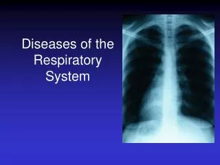
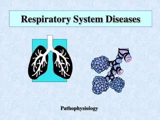
PATHOPHYSIOLOGY OF RESPIRATORY SYSTEM DISEASES. Mehtap KAÇAR KOÇAK M.D. PhD Yeditepe University Medicine School, Pathophysiology. General Introduction. Respiratory diseases is often classified as acute or chronic, obstructive or restrictive, and infectious or noninfectious.

E N D
PATHOPHYSIOLOGY OF RESPIRATORY SYSTEM DISEASES • Mehtap KAÇAR KOÇAK M.D. PhD Yeditepe University Medicine School, Pathophysiology
General Introduction • Respiratory diseases is often classified as acute or chronic, obstructive or restrictive, and infectious or noninfectious.
Reduction of Pulmonary Function • Inadequate blood flow to the lungs – hypoperfusion • Inadequate air flow to the alveoli - hypoventilation
Signs and Symptoms of Pulmonary Disease • Dyspnea, • Abnormal breathing patterns, • Hypoventilation and hyperventilation, • Cough, • Hemoptysis, • Cyanosis, • Pain, • Clubbing, • Abnormal sputum.
Dyspnea • Dyspnea – subjective sensation of uncomfortable breathing, feeling “short of breath” • Ranges from mild discomfort after exertion to extreme difficulty breathing at rest. • Usually caused by diffuse and extensive rather than focal pulmonary disease.
Dyspnea • Due to: • Airway obstruction • Greater force needed to provide adequate ventilation • Wheezing sound due to air being forced through airways narrowed due to constriction or fluid accumulation • Decreased compliance of lung tissue
Signs of dyspnea: • Flaring nostrils • Use of accessory muscles in breathing • Retraction (pulling back) of intercostal spaces
Types of dyspnea • Orthopnea; is caused by the horizontal position, which redistributes body water, causes the abdominal contents to exert pressure on the diaphragm, and decreases the efficiency of the respiratory muscles. • Some individuals with left ventricular failure wake up at night gasping for air and must sit up or stand to relieve the dispnea, this type of positional dyspnea is termed Paroxysmal nocturnal dyspnea (PND) • PND results from fluid in the lungs caused by the redistribution of of body water while the individual is recumbent.
Abnormal breathing patterns • Normal breathing (eupnea) is rhythmic and effortless. Ventilatory rate is 8 to 16 breaths per minute, and tidal volume ranges from 400 to 800 ml. • The rate, depht, regulatory and effort of breathing undergo characteristic alterations in response to physiologic and pathophysiologic conditions…..
Kussmaul respiration (hyperpnea); is characterized by →a slightly increased ventilatory rate, → very large tidal volume, → no expiration pause. • Strenous exercise or metabolic acidosis induces Kussmaul respiration. • Labored or obstructed breathing consists of slow ventilatory rate, large tidal volume, increased effort, and prolonged inspiration or expiration, depending on the site of obstruction. • Audible wheezing (whistling sounds) and stridor (high-pitched sounds made during inspiration) is often present.
Restricted breathing is characterized by small tidal volumes and rapid ventilatory rate (tachypnea). • Restricted breathing is commonly caused by disorders such as pulmonary fibrosis that stiffen the lungs or chest wall and decrease compliance. • Panting occurs with exercise. • Shock and severe cerebral hypoxia contribute to gasping respirations that consist of irregular, quick inspirations with an expiratory pause.
Sighing respirations consists of irregular breathing characterized by frequent, deep sighing inspirations. Sighing respirations are caused by anxiety. • Cheyne-Stokes respirations are characterized by alternating periods of deep and shallow breathing. Apnea lasting 15 to 60 seconds is followed by ventilations that increase in volume until a peak is reached, after which ventilation (tidal volume) decreases again to apnea. • This respirations results from any condition that slows the blood flow to the brain stem, which in turn slows impulses sending information to the respiratory centers of the brain stem.
Hypoventilation and hyperventilation • Hypoventilation is inadequate alveolar ventilation in relation to metabolic demands. It is caused by alterations in pulmonary mechanics or in the neurologic control of breathing. • With hypoventilation, CO2 removal does not keep up with CO2 production and PaCO2 increases, causing hypercapnia (PaCO2>44 mmHg). • This results in respiratory acidosis, which can affect the function of many tissues throughout the body. (somnolence or disorientation)
Hyperventilation is alveolar ventilation that exceeds metabolic demands. The lungs remove CO2 at a faster rate than it is produced by cellular metabolism, resulting in decreased PaCO2 or hypocapnia (PaCO2<36 mmHg). • Hyperventilation results in a respiratory alkalosis that also can interfere with tissue function. • Hyperventilation commonly occurs with severe anxiety, acute head injury, and conditions that cause insufficient oxygenation of the blood. • Hypoventilation and hyperventilation can be determined only by arterial blood gas analysis.
Blood Gas Analysis • Factors Connected with blood gas measurements • Abnormalities of respiratory control • Gas exchange • Respiratory mechanics • Circulation
Blood Gas Analysis • Blood gas values • pH : 7.35—7.45 • PaO2: 80—100mmHg(10.6~13.3kpa) • SaO2: 91—97.7% • PaCO2: 35—45mmHg(4.67~6.0kpa)
Cough • Attempt to clear the lower respiratory passages by abrupt and forceful expulsion of air • Most common when fluid accumulates in lower airways • Most coughs are initiated in the larynx and in the tracheabronchial tree by both mechanical and chemical irritant receptor stimulation.
Other cough receptors are located: • The external auditory canal, • Diaphragm, • Pericardium, • Pleura, • Stomach, • The most distal bronchioli and the alveoli (a few). • Stimulation of cough receptors is transmitted centrally through the vagus nerve. (this way is inhibited by opiates and serotoninergic agents)
Acute cough: • It is cough that resolves within 2 to 3 weeks of the onset of illness or resolves with treatment of the underlying condition. • It is commonly the result of; • Upper respiratory infections, • Allergic rhinitis, • Acute bronchitis, • Pneumonia, • Congestive heart failure, • Pulmonary embolus, • Aspiration.
Chronic cough: • It is defined as cough that has persisted for more than 3 weeks. • In nonsmokers, chronic cough is almost always caused by • postnasal drainage syndrome, • Asthma, • Gastroesophageal reflux disease. • In smokers, chronic bronchitis is the most common cause of chronic cough.
Hemoptysis: • Hemoptysis is the coughing up of blood or bloody secretion. • Hemoptysis is sometimes confused with hematemesis, which is the vomiting of blood. • Blood that is coughed up is usually bright red, has an alkaline pH, and is mixed frothy sputum, whereas blood that is vomited is dark, has an acidic pH, and is mixed with food particles.
Hemoptysis results from damage to the lung parenchyma with rupture of pulmonary vessels or from inflammation, injury, or cancer of the bronchial tree.
The most common causes of hemoptysis are: • Tuberculosis, • Bronchiectasis, • Lung cancer, • Bronchitis, • Pneumonia.
Cyanosis • Cyanosis is bluish discoloration of the skin and mucous membranes caused by increasing amounts of desaturated or reduced hemoglobin (which is bluish) in the blood. • Cyanosis generally develops when 5 g of hemoglobin is desaturated, regardless of hemoglobin concentration.
Cyanosis can be caused by decreased arterial oxygenation (low PaO2), pulmonary or cardiac right-to-left shunts, decreased cardiac output, cold environments, or anxiety. • Lack of cyanosis does not necessarily indicate that oxygenation is normal. For example, severe anemia and CO poisoning can cause inadequate oxygenation of tissues without causing cyanosis.
Most cases arise as a result of peripheral vasoconstriction – result is reduced blood flow, which allows hemoglobin to give up more of its oxygen to tissues- peripheral cyanosis. • Best seen in nail beds • Due to cold environment, anxiety, etc.
Central cyanosis can be due to : • Abnormalities of the respiratory membrane • Mismatch between air flow and blood flow • Expressed as a ratio of change in ventilation (V) to perfusion (Q) : V/Q ratio • Pulmonary thromboembolus - reduced blood flow • Airway obstruction – reduced ventilation • In persons with dark skin can be seen in the whites of the eyes and mucous membranes.
Pain • Pain caused by pulmonary disorders originates in the pleurae, airways or chest wall. • Pleural pain is the most common pain caused by pulmonary disease and is usually sharp or stabbing in character. (infection and inflammation) • Pleural pain is also common with pulmonary infarction caused by pulmonary embolism.
Pulmonary pain is central chest pain that is pronounced after coughing and occurs in individulas with infection and inflammation of the trachea or bronchi. (it is differentiated from cardiac pain). • Pain in the chest wall is muscle pain or rib pain. (excessive coughing, costochondritis)
Clubbing • Clubbing is the selective bulbous enlargement of the end (distal segment) of a digit (finger or toe), whose severity can be graded from 1 to 5 based on the extend of nail bed hypertrophy and the amount of changes in the nails themselves.
Clubbing is commonly associated with diseases that interfere with oxygenation, such as bronchiectasis, cystic fibrosis, pulmonary fibrosis, lung abscess and congenital heart disease. • Lung cancer is sometimes associated with clubbing even in the absence of significant hypoxemia. • This syndrome is called hypertrophic osteoarthropathy (HOA) and its pathogenesis is unknown.
Abnormal sputum • The color, consistency, odor and amount of sputum vary different pulmonary disorders.
Conditions Caused by Pulmonary Disease or Injury • Hypercapnia, • Hypoxemia, • Acute respiratory failure, • Pulmonary edema, • Aspiration, • Atelectasis, • Bronchiectasis, • Bronchiolitis, • Pleural abnormalities (pneumothorax, pleural effusion, empyema) • Abscess formation and cavitation, • Pulmonary fibrosis
Hypercapnia • Hypercapnia or increased CO2 in the arterial blood is caused by hypoventilation of the alveoli. • Causes include; • * depression of the respiratory center by drug, • * infection of CNS or trauma, • * spinal cord disruption or poliomyelitis, • * diseases of the neuromuscular junction (MG, MD) • * chest injury, • * large airway obstruction. • Hypercapnia and the associated respiratory acidosis can result in several important clinical manifestations. (dysrythmia, coma, somnolence)
Hypoxemia • Hypoxemia or reduced oxygenation of arterial blood is caused by respiratory alterations. • The five causes of hypoxemia are; • * decreased oxygen content in inspired gas (high altitude), • * hypoventilation (drug overdose, neurologic damage), • * diffusion abnormalities (emphysema, fibrosis, edema), • * abnormal ventilation-perfusion ratios (asthma, chronic bronchitis, ARDS), • * pulmonary right-to-left shunt (congenital heart defects). • Hypoxemia is often associated with a compensatory hyperventilation and resultant respiratory alkalosis. • Hypoxemia result in widwspread tissue dysfunction and when severe can lead to organ infarction.
Adult Respiratory Pathophysiology McCance & Heuther 32.02 W. Rose NURSING 621
Acute respiratory failure • Respiratory failure is defined as inadequate gas exchange, that is hypoxemia, where • PaO2 is ≤50 mmHg • PaCO2 is ≥50mmHg, (hypercapnia) • pH ≤7.25.
Pulmonary edema • Pulmonary edema is excess water in the lung. • The normal lung contains very little water or fluid. • It is kept dry by lymphatic drainage and a balance among capillary hydrostatic pressure, capillary oncotic pressure and capillary permeability. • The most common cause of pulmonary edema is heart disease.
Pulmonary Circulation: • Consists of: • Pulmonary artery & its branches • Thin-walled, easily distensible Pulmonary capillaries • Pulmonary venous system • Left Atrium • Low pressure • Low resistance to blood flow • Accepts all blood from right heart
PA pressure: Sys: 20 – 30 mm Hg Dias: 8 – 12 mmHg. 12 – 15 mm Hg mean Normal L.A.Pr: 8 mm Hg with an Upper limit of 15 mm Hg Blood vol: About 1 litre. More than 100 ml in capillaries.
Pulmonary Circulation • Pul cap pr.: about 10 mm Hg • Oncotic pressure: 25 mm Hg • Inward gradient: 15 mm Hg • Pul congestion and oedema result when the pul cap pr is >25 mm Hg (“backward failure”)
Pulmonary Arterial Pressure rises during: • Negative pressure breathing • Supine position • Systemic venous congestion from any cause • Overtransfusion • Left ventricular failure
Pulmonary Arterial Pressurefalls during: • Positive pressure breathing • Upright position • Valsalva manuevre • After haemorrhage • Systemic vasodilatation from any cause.
PULMONARY EDEMA Classified into: Cardiogenic: Otherwise called as Hydrostatic or Transudative. Non-Cardiogenic: Other-wise called as Permeability or Exudative.
Noncardiogenic Pulmonary Edema Cardiogenic Pulmonary Edema • Increased pulmonary vascular pressure • Transudation of fluid across endothelium into pulmonary interstitium and then into the alveolar space. • Low pressure pulmonary edema • Injured microvascular endothelium allows protein-rich fluid to enter theextravascular spaces.
Cardiogenic Pulmonary Edema Causes: Noncardiogenic Pulmonary Edema Causes: • Left ventricular failure • Volume overload • Mechanical obstruction of left outflow tract e.g. Mitral stenosis • Other valvular diseases like Mitral stenosis, PDA, Aortic valvular diseases & also in congestive failure and hypertension. • Toxins: eg. Smoke, ozone, phosgene, Nitrogen dioxide, • Emboli: Air, thrombotic, amniotic fluid • Trauma and burns • Aspiration of gastric contents • Acute radiation Pneumonitis • D.I.C.
PULMONARY EDEMA MIXED PATHOGENESIS • Incompletely understood: • High altitude pulmonary edema • Neurogenic: midbrain lesions, Middle/Posterior cranial fossa surgeries • Reperfusion pulmonary edema • Narcotic overdose • Tocolytic therapy • Uraemia • Negative pressure pulmonary edema.
Adult Respiratory Pathophysiology McCance & Heuther 32.03 W. Rose NURSING 621
PUL EDEMA – SIGNS & SYMPTOMS Symptoms: Acute Dyspnea Cough, Pink frothy sputum Signs: Tachycardia Blood Pressure: variable Lung crepitations