Download
1 / 21
210 likes | 224 Views
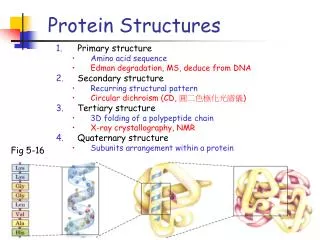
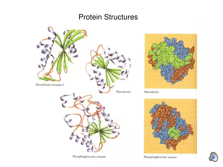
Protein Structures. Ramachandran plot. Shows grouping of φψ combinations and relates them to structures in real proteins Repetitive structures ( - helices, -sheets) are common. -helix. • 3.6 amino acids per turn • 0.54 nm per turn • side chains pointed out

E N D
Ramachandran plot Shows grouping of φψ combinations and relates them to structures in real proteins Repetitive structures (- helices, -sheets) are common.
-helix • 3.6 amino acids per turn • 0.54 nm per turn • side chains pointed out • H-bonds parallel to axis • n-4 H-bonds • dipole moment (neg. at C end) • no pro, less gly, ser • limited similar side chain charges
-helices have a dipole moment some side chains are preferred
ß-sheets are parallel or anti-parallel
ß-sheets can form a “ß-barrel” A recent paper elucidates the ß-barrel structure of a toxic amyloid protein Laganowsky et al., “Atomic view of a toxic amyloid small oligomer”, Science 9 March 2012, 335:1228
A reverse turn (ß-bend): R2 (C=O side) is often G,A R3 (N-H side) is often D Proline is often R2 or R3
Tertiary structure involves bonds between and among side chains: • Hydrogen (-O-H…O-) • Ionic (generally repulsion: -CH2-NH3+:::::::+H3N-CH2-) • Van der Waal’s (short distance attraction) • Disulfide (covalent: -CH2-S-S-CH2-) • Hydrophobic
The types of side chains, and the tertiary bonds they form, influence the positions of secondary structures.
And the position of a secondary structure in a protein will influence the types of side chains (tertiary structure). An -helix on the surface of a protein will have hydrophilic side chains on one side of the helix axis and hydrophobic side chains on the other.
An -helix in the interior of a protein will have primarily hydrophobic side chains.
An -helix exposed to the solution on all sides (unusual) will have hydrophilic side chains on all sides of the helix axis (mostly).
Quaternary structures Involve separate polypeptides held together with weak bonds in various symmetries Symmetries Homomultimer:: heteromultimer Isologous:: heterologous Closed::open E.g.: tubulin, actin, TMV coat E.g.: hemoglobin
Secondary-tertiary structure of UVR8 subunits involves multiple ß-sheets. Quaternary structure involves electrostatic interactions between positively charged arginines and negatively charged aspartates.
The folding of a protein reduces the free energy (G) of the system. Folding states
The folding of a protein involves both protein and solvent. • G = GF- GU • = H - TS • = • + H(protein) • + H(solvent) • - TS(protein) • - TS(solvent) • G for folding is small (-20 to -60 kJ/mol) and primarily from hydrophobic interactions • Why so low?
Changes in shape are an important part of protein function and control. For example: a change in shape allows DNA methyltransferase to choose hemi-methylated meCG/GC for bimethylation to meCG/GmeC Science 25 Feb 2011: Song, et al., 331:1036
Summary: Primary structure involves bonds between amino and carboxylic groups, stabilizing the amino acid sequence Secondary structure involves hydrogen bonds between back- bone atoms, forming -helices, ß-sheets, and ß-bends. Tertiary structure involves bonds between side chains. Quaternary structure involves bonds connecting separate poly- peptide chains. G for folding is small and primarily from hydrophobic interactions. Stigler et al., The complex folding network of single calmodulin molecules. Science 334:512, 28 October 2011 Lindorff-Larsen et al., How fast-folding proteins fold. Science 334:517, 28 October 2011 Dill and MacCallum, The protein-folding problem, 50 years on. Science 338:1042, 23 November 2012 Saibil, Machinery to reverse irreversible aggregates. Science 339:1040, 1080 March 2013