Download
1 / 32
320 likes | 323 Views
Learn about the stages of mitosis, the eukaryotic cell cycle, and how cytokinesis occurs. Understand the process of cell division and the organization of chromosomes during mitosis.

E N D
Section 2: Mitosis Preview • Bellringer • Key Ideas • Eukaryotic Cell Cycle • Stages of Mitosis • Cytokinesis • Summary
Bellringer Using the diagram of a football field, write a short paragraph describing how the players, the midfield line, and the goal posts compare to the structures of a cell involved in mitosis and cell division.
Key Ideas • What are the phases of the eukaryotic cell cycle? • What are the four stages of mitosis? • How does cytokinesis occur?
Eukaryotic Cell Cycle • The cell cycle is a repeating sequence of cellular growth and division during the life of a cell. • The life of a eukaryotic cell cycles through phases of growth, DNA replication, preparation for cell division, and division of the nucleus and cytoplasm. • The cell cycle is made up of five phases. The first three phases together are known as interphase. The remaining two phases make up cell division.
Eukaryotic Cell Cycle, continued Interphase • During interphase, the cell is not dividing. It is growing and preparing to divide. • Different types of cells spend different amounts of time in interphase. • Cells that divide often, such as skin cells, spend less time in interphase. Cells that divide seldom, such as nerve cells, spend most of their time in interphase.
Eukaryotic Cell Cycle, continued Interphase • During the first gap phase (G1), a cell grows rapidly as the cell builds more organelles. For most organisms, this phase occupies the major portion of the cell’s life. • During the synthesis phase (S), a cell’s DNA is copied. At the end of the S phase, the cell’s nucleus has twice as much DNA as it did in the G1 phase. • During the second gap phase (G2), the cell continues to grow and prepares to divide. Hollow protein fibers called microtubules are organized in the cytoplasm during G2.
Visual Concept: Cell Cycle—G1 Phase Click above to play the video.
Visual Concept: Cell Cycle—S Phase Click above to play the video.
Visual Concept: Cell Cycle—G2 Phase Click above to play the video.
Visual Concept: Cell Cycle—M Phase Click above to play the video.
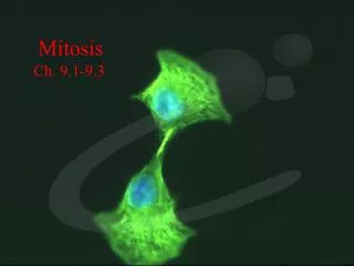
Eukaryotic Cell Cycle, continued Cell Division • Each new cell requires a complete set of organelles, including a nucleus. • The process of dividing the nucleus into two daughter nuclei is called mitosis. • The process of separating the organelles and the cytoplasm is called cytokinesis.
Visual Concept: Mitosis Click above to play the video
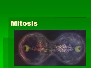
Eukaryotic Cell Cycle, continued Cell Division • During mitosis, the nucleus divides to form two nuclei. Each nucleus contains a complete set of the cell’s chromosomes. • The nuclear membrane breaks down briefly. The two sister chromatids of each chromosome are pulled to the opposite sides of the dividing cell.
Eukaryotic Cell Cycle, continued Cell Division • As the nucleus divides, the cytoplasm also begins to divide. • Each daughter cell receives about half of the original cell’s organelles. • During cytokinesis, the two daughter cells are physically separated.
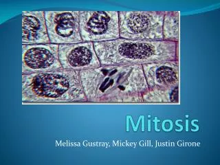
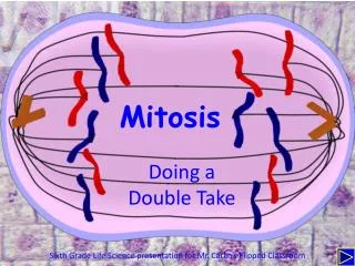
Stages of Mitosis • Although mitosis is a continuous process, biologists traditionally divide it into four stages. • Mitosis is a continuous process that can be observed in four stages: prophase, metaphase, anaphase, and telophase.
Stages of Mitosis, continued Stage 1 Prophase • Within the nucleus, chromosomes begin to condense and become visible under a light microscope. • The nuclear membrane breaks down. Outside the nucleus, a special structure called the spindle forms. The spindle is made up of several spindle fibers. • Each spindle fiber in turn is made up of an individual microtubule—a hollow tube of protein. Microtubules organize into a spindle that runs at a right angle to the cell’s equator.
Stages of Mitosis, continued Stage 1 Prophase • Cells have an organelle called the centrosome, which helps assemble the spindle. • In animal cells, the centrosome includes a pair of centrioles. Each centriole is made up of nine triplets of microtubules arranged as a short, hollow tube. • Before mitosis, the cell’s centrosome is duplicated. During prophase, the centrosomes move to opposite poles of the cell.
Prophase Click to animate the image. C B F E A D
Stages of Mitosis, continued Stage 2 Metaphase • During metaphase, the chromosomes are packaged into their most condensed form. • The nuclear membrane is fully dissolved, and the condensed chromosomes move to the center of the cell and line up along the cell’s equator. • Spindle fibers form a link between the poles and the centromere of each chromosome.
Stages of Mitosis, continued Stage 3 Anaphase • Once all of the chromosomes are lined up, the spindle fibers shorten. The spindle fibers shorten by breaking down the microtubules bit by bit. • Sister chromatids move toward opposite poles as the spindle fibers that are attached continue to shorten. • Each pole now has a full set of chromosomes.
Stages of Mitosis, continued Stage 4 Telophase • A nuclear envelope forms around the chromosomes at each pole of the cell. • Chromosomes, now at opposite poles, uncoil and change back to their original chromatin form. • The spindle dissolves and the spindle fibers break down and disappear. • Mitosis is complete.
Cytokinesis • As mitosis ends, cytokinesis begins. The cytoplasm is separated, and two cells are formed. • During cytokinesis, the cell membrane grows into the center of the cell and divides it into two daughter cells of equal size. • Each daughter cell has about half of the parent’s cytoplasm and organelles. • The end result of mitosis and cytokinesis is two genetically identical cells in place of the original cell.
Cytokinesis, continued Separating the Cytoplasm • In animal cells and other cells that lack cell walls, the cell is pinched in half by a belt of protein threads. • In plant cells and other cells that have rigid cell walls, the cytoplasm is divided in a different way.
Cytokinesis, continued Separating the Cytoplasm • Vesicles holding cell wall material line up across the middle of the cell. • These vesicles fuse to form a large, membrane-bound cell wall called the cell plate. • When it is completely formed, the cell plate separates the plant cell into two new plant cells.
Visual Concept: Comparing Cell Division in Plants and Animals
Cytokinesis, continued Continuing the Cell Cycle • After cytokinesis is complete, each cell enters the G1 stage of interphase. • The daughter cells are about equal in size—about half the size of the original cell. • The activity of each cell continues because each has its own DNA and organelles. The cell cycle continues for each new cell.
Summary • The life of a eukaryotic cell cycles through phases of growth, DNA replication, preparation for cell division, and division of the nucleus and cytoplasm. • Mitosis is a continuous process that can be observed in four stages: prophase, metaphase, anaphase, and telophase. • During cytokinesis, the cell membrane grows into the center of the cell and divides it into two daughter cells of equal size. Each daughter cell has about half of the parent’s cytoplasm and organelles.