Download
1 / 55
550 likes | 670 Views
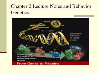
Genetics Lecture 2 Mitosis. DNA. Every living thing contains a substance described as the genetic material. Except in certain viruses, this material is composed of the nucleic acid, DNA.

E N D
DNA • Every living thing contains a substance described as the genetic material. • Except in certain viruses, this material is composed of the nucleic acid, DNA. • DNA has an underlying linear structure possessing segments called genes, the products of which direct the metabolic activities of cells.
An organisms DNA, with its arrays of genes, is organized into structures called chromosomes, which serve as vehicles for transmitting genetic information. • The manner in which chromosomes are transmitted from one generation of cells to the next and from organisms to their descendants must be exceedingly precise.
mitosis and meiosis • Two major processes are involved in the genetic continuity of nucleated cells: mitosis and meiosis. • Although the mechanisms of the two processes are similar in many ways, the outcomes are quite different. • Mitosis leads to the production of two cells, each with the same number of chromosomes as the parent cell. • In contrast, meiosis reduces the genetic content and the number of chromosomes by precisely half
This reduction is essential if sexual reproduction is to occur without doubling the amount of genetic material in each new generation. • Strictly speaking, mitosis is that portion of the cell cycle during which the hereditary components are equally partitioned into daughter cells. • Meiosis is part of a special type of cell division that leads to the production of sex cells: gametes or spores. • This process is an essential step in the transmission of genetic information from an organism to its offspring.
Normally, chromosomes are visible only during mitosis and meiosis. • When cells are not undergoing division, the genetic material making up chromosomes unfolds and uncoils into a diffuse network within the nucleus, generally referred to as chromatin. • Before describing mitosis and meiosis, we will briefly review the structure of cells, emphasizing components that are of particular significance to genetic function. • We will also compare the structural differences between the prokaryotic (non-nucleated) cells of bacteria and the eukaryotic cells of higher organisms.
Cell Structure Is Closely Tied to Genetic Function • Before 1940, our knowledge of cell structure was limited to what we could see with the light microscope. • Around 1940, the transmission electron microscope was in its early stages of development, and by 1950, many details of cell ultrastructure had emerged. • Under the electron microscope, cells were seen as highly varied, highly organized structures whose form and function are dependent on specific genetic expression by each cell type.
A new world of whorled membranes, organelles, microtubules, granules, and filaments was revealed • These discoveries revolutionized thinking in the entire field of biology. • Many cell components, such as the nucleolus, ribosome, and centriole, are involved directly or indirectly with genetic processes. • Other components- the mitochondria and chloroplasts-contain their own unique genetic information.
Two major groups • Living organisms are categorized into two major groups depending on whether or not their cells contain a nucleus. • The presence of a nucleus and other membranous organelles is the defining characteristic of eukaryotic organisms. • The nucleus in eukaryotic cells is a membrane-bound structure that houses the genetic material, DNA, which is complexed with an array of acidic and basic proteins into thin fibers. • During non-divisional phases of the cell cycle, the fibers are uncoiled and dispersed into chromatin.
During mitosis and meiosis, chromatin fibers coil and condense into chromosomes. • Also present in the nucleus is the nucleolus, an amorphous component where ribosomal RNA (rRNA) is synthesized and where the initial stages of ribosomal assembly occur. • The portions of DNA that encode rRNA are collectively referred to as the nucleolus organizer region, or the NOR.
Prokaryotic organisms • Prokaryotic organisms, of which there are two major groups, lack a nuclear envelope and membranous organelles. • For the purpose of our brief discussion here, we will consider the eubacteria, the other group being the more ancient bacteria referred to as archaea
In eubacteria, such as Escherichia coli, the genetic material is present as a long, circular DNA molecule that is compacted into an unenclosed region called the nucleoid. • Although the DNA is compacted, it does not undergo the extensive coiling characteristic of the stages of mitosis, during which the chromosomes of eukaryotes become visible.
eukaryotic cell • The remainder of the eukaryotic cell within the plasma membrane, excluding the nucleus, is referred to as cytoplasm and includes a variety of extranuclear cellular organelles. • In the cytoplasm, a nonparticulate, colloidal material referred to as the cytosol surrounds and encompasses the cellular organelles.
The cytoplasm also includes an extensive system of tubules and filaments, comprising the cytoskeleton, which provides a lattice of support structures within the cell. • Consisting primarily of microtubules, which are made of the protein tubulin, and microfilaments, which derive from the protein actin, this structural framework maintains cell shape, facilitates cell mobility and anchors the various organelles.
One organelle, the membranous endoplasmic reticulum (ER), compartmentalizes the cytoplasm, greatly increasing the surface area available for biochemical synthesis. • The ER appears smooth in places where it serves as the site for synthesizing fatty acids and phospholipids; in other places, it appears rough because it is studded with ribosomes. • Ribosomes serve as sites where genetic information contained in messenger RNA (mRNA) is translated into proteins.
other cytoplasmic structures • Three other cytoplasmic structures are very important in the eukaryotic cell’s activities: mitochondria, chloroplasts, and centrioles. • Mitochondria are found in most eukaryotes, including both animal and plant cells, and are the sites of the oxidative phases of cell respiration.
These chemical reactions generate large amounts of the energy-rich molecule adenosine triphosphate (ATP). • Chloroplasts, which are found in plants, algae, and some protozoans, are associated with photosynthesis, the major energy-trapping process on Earth.
Both mitochondria and chloroplasts contain DNA in a form distinct from that found in the nucleus. • They are able to duplicate themselves and transcribe and translate their own genetic information. • It is interesting to note that the genetic machinery of mitochondria and chloroplasts closely resembles that of prokaryotic cells. • This and other observations have led to the proposal that these organelles were once primitive free- living organisms that established symbiotic relationships with primitive eukaryotic cells.
This theory concerning the evolutionary origin of these organelles is called the endosymbiont hypothesis. • Animal cells and some plant cells also contain a pair of complex structures called centrioles. • These cytoplasmic bodies, located in a specialized region called the centrosome, are associated with the organization of spindle fibers that function in mitosis and meiosis.
In some organisms, the centriole is derived from another structure, the basal body, which is associated with the formation of cilia and flagella (hair-like and whip-like structures for propelling cells or moving materials). • Over the years, many reports have suggested that centrioles and basal bodies contain DNA, which could be involved in the replication of these structures. This idea is still being investigated.
The organization of spindle fibers by the centrioles occurs during the early phases of mitosis and meiosis. • These fibers play an important role in the movement of chromosomes as they separate during cell division. • They are composed of arrays of microtubules consisting of polymers of the protein tubulin.
Chromosomes Exist in Homologous Pairs in Diploid Organisms • As we discuss the processes of mitosis and meiosis, it is important to understand the concept of homologous chromosomes. • Chromosomes are most easily visualized during mitosis. • When they are examined carefully, distinctive lengths and shapes are apparent. • Each chromosome contains a constricted region called the centromere, whose location establishes the general appearance of each chromosome. Figure 2-3 shows chromosomes with centromere placements at different distances along their length.
Extending from either side of the centromere are the arms of the chromosome. • Depending on the position of the centromere, different arm ratios are produced. • As Figure 2-3 illustrates, chromosomes are classified as metacentric, submetacentric, acrocentric, or telocentric on the basis of the centromere location. • The shorter arm, by convention, is shown above the centromere and is called the p arm (p, for “petite”). • The longer arm is shown below the centromere and is called the q arm (q because it is the next letter in the alphabet).
mitosis • In the study of mitosis, several other observations are of particular relevance. • First, all somatic cells derived from members of the same species contain an identical number of chromosomes. • In most cases, this represents the diploid number (2n), • When the lengths and centromere placements of all such chromosomes are examined, a second general feature is apparent. • With the exception of sex chromosomes, they exist in pairs with regard to these two properties, and the members of each pair are called homologous chromosomes. • So, for each chromosome exhibiting a specific length and centromere placement, another exists with identical features
There, the human mitotic chromosomes have been photographed, cut out of the print, and matched up, creating a display called a karyotype. • humans have a 2n number of 46 chromosomes, which on close examination exhibit a diversity of sizes and centromere placements. • Note also that each of the 46 chromosomes in this karyotype is clearly a double structure consisting of two parallel sister chromatids connected by a common centromere. • Had these chromosomes been allowed to continue dividing, the sister chromatids, which are replicas of one another, would have separated into the two new cells as division continued.
The haploid number (n) of chromosomes is equal to one-half the diploid number. • Collectively the genetic information contained in a haploid set of chromosomes constitutes the genome of the species. • This, of course, includes copies of all genes as well as a large amount of noncoding DNA.
Homologous chromosomes • Homologous chromosomes have important genetic similarities. • They contain identical gene sites along their lengths; each site is called a locus (pl. loci). • Thus, they are identical in the traits that they influence and in their genetic potential. • In sexually reproducing organisms, one member of each pair is derived from the maternal parent (through the ovum) and the other member is derived from the paternal parent (through the sperm).
Therefore, each diploid organism contains two copies of each gene as a consequence of biparental inheritance, inheritance from two parents. • The members of each pair of genes, while influencing the same characteristic or trait, need not be identical. • In a population of members of the same species, many different alternative forms of the same gene, called alleles, can exist.
The concepts of haploid number, diploid number, and homologous chromosomes are important for understanding the process of meiosis. • During the formation of gametes or spores, meiosis converts the diploid number of chromosomes to the haploid number. • As a result, haploid gametes or spores contain precisely one member of each homologous pair of chromosomes—that is, one complete haploid set. • Following fusion of two gametes at fertilization, the diploid number is reestablished; that is, the zygote contains two complete haploid sets of chromosomes. • The constancy of genetic material is thus maintained from generation to generation.
sex • There is one important exception to the concept of homologous pairs of chromosomes. • ln many species, one pair, consisting of the sex-determining chromosomes, is often not homologous in size, centromere placement, arm ratio, or genetic content. • For example, in humans, while females carry two homologous X chromosomes, males carry one Y chromosome in addition to one X chromosome • These X and Y chromosomes are not strictly homologous. • The Y is considerably smaller and lacks most of the gene loci contained on the X. • Nevertheless, they contain homologous regions and behave as homologs in meiosis so that gametes produced by males receive either one X or one Y chromosome.
Mitosis Partitions Chromosomes into Dividing Cells • The process of mitosis is critical to all eukaryotic organisms. • In some single-celled organisms, such as protozoans and some fungi and algae, mitosis (as a part of cell division) provides the basis for asexual reproduction. • Multicellular diploid organisms begin life as single-celled fertilized eggs called zygotes. • The mitotic activity of the zygote and the subsequent daughter cells is the foundation for the development and growth of the organism. • In adult organisms, mitotic activity is the basis for wound healing and other forms of cell replacement in certain tissues.
For example, the epidermal cells of the skin and the intestinal lining of humans are continuously sloughed off and replaced. • Cell division also results in the continuous production of reticulocytes that eventually shed their nuclei and replenish the supply of red blood cells in vertebrates. • In abnormal situations, somatic cells may lose control of cell division, and form a tumor.
The genetic material is partitioned into daughter cells during nuclear division, or karyokinesis. This process is quite complex and requires great precision. • The chromosomes must first be exactly replicated and then accurately partitioned. The end result is the production of two daughter nuclei, each with a chromosome composition identical to that of the parent cell.
Karyokinesis • Karyokinesis is followed by cytoplasmic division, or cytokinesis. • This less complex process requires a mechanism that partitions the volume into two parts, and then encloses each new cell in a distinct plasma membrane. • As the cytoplasm is reconstituted, organelles either replicate themselves, arise from existing membrane structures, or are synthesized de novo (anew) in each cell. • Following cell division, the initial size of each new daughter cell is approximately one-half the size of the parent cell. • However, the nucleus of each new cell is not appreciably smaller than the nucleus of the original cell. • Quantitative measurements of DNA confirm that there is an amount of genetic material in the daughter nuclei equivalent to that in the parent cell.
Interphase and the Cell Cycle • Many cells undergo a continuous alternation between division and non-division. • The events that occur from the completion of one division until the completion of the next division constitute the cell cycle (Figure 2-5). • We will consider interphase, the initial stage of the cell cycle, as the interval between divisions. • It was once thought that the biochemical activity during interphase was devoted solely to the cell’s growth and its normal function.
However, we now know that another biochemical step critical to the ensuing mitosis occurs during interphase: the replication of the DNA of each chromosome. • This period, during which DNA is synthesized, occurs before the cell enters mitosis and is called the S phase. • The initiation and completion of synthesis can be detected by monitoring the incorporation of radioactive precursors into DNA.
Investigations of this nature demonstrate two periods during interphase when no DNA synthesis occurs, one before and one after the S phase. • These are designated G1 (gap I) and G2 (gap II), respectively • During both of these intervals, as well as during S, intensive metabolic activity, cell growth, and cell differentiation are evident. • By the end of G2, the volume of the cell has roughly doubled, DNA has been replicated, and mitosis (M) is initiated. • Following mitosis, continuously dividing cells then repeat this cycle (G1, S, G2, M) over and over, as shown in Figure 2—5.
G1 is of great interest in the study of cell proliferation and its control. • At a point during G1, all cells follow one of two paths. • They either withdraw from the cycle, become quiescent, and enter the G0 stage), or they become committed to proceed through G1, initiating DNA synthesis, and completing the cycle. • Cells that enter GO remain viable and metabolically active but are not proliferative. • Cancer cells apparently avoid entering G0 or pass through it very quickly. • Other cells enter G0 and never reenter the cell cycle. • Still other cells in GO can be stimulated to return to G1 and thereby reenter the cell cycle.
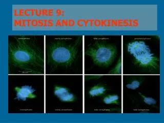
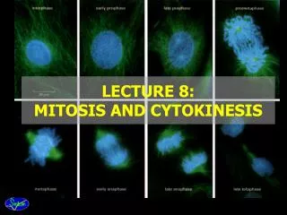
Prophase • Often, over half of mitosis is spent in prophase, a stage characterized by several significant occurrences. • One of the early events in prophase of all animal cells is migration of two pairs of centrioles to opposite ends of the cell. • These structures are found just outside the nuclear envelope in an area of differentiated cytoplasm called the centrosome. • It is believed that each pair of centrioles consists of one mature unit and a smaller, newly formed centriole.
Prometaphase and Metaphase • The distinguishing event of the two ensuing stages is the migration of every chromosome, led by its centromeric region, to the equatorial plane. • The equatorial plane, also referred to as the metaphase plate, is the midline region of the cell, a plane that lies perpendicular to the axis established by die spindle fibers. • In some descriptions, the term prometaphase refers to the period of chromosome movement [Figure 2-7(c)], and the term metaphase is applied strictly to the chromosome configuration following migration.
Migration • Migration is made possible by the binding of spindle fibers to the chromosomes kinetochore, an assembly of multilayered plates of proteins associated with the centromere. • This structure forms on opposite sides of each paired centromere, in intimate association with the two sister chromatids. • Once properly attached to the spindle fibers, cohesin is degraded by an enzyme, appropriately named separase, and the sister chromatid arms disjoin, except at the centromere region.
A unique protein family called shugoshin (from the Japanese meaning guardian spirit) protects cohesin from being degraded by separase at the centromeric regions. • The involvement of the cohesin and shugoshin complexes with a pair of sister chromatids during mitosis is depicted in Figure 2-8. • At the completion of metaphase, each centromere is aligned at the metaphase plate with the chromosome arms extending outward in a random array
Anaphase • Events critical to chromosome distribution during mitosis occur during anaphase, the shortest stage of mitosis. • During this phase, sister chromatids of each chromosome, held together only at their centromere regions, disjoin (separate) from one another—an event described as disjunction—and are pulled to opposite ends of the cell.