Download
1 / 13
130 likes | 248 Views
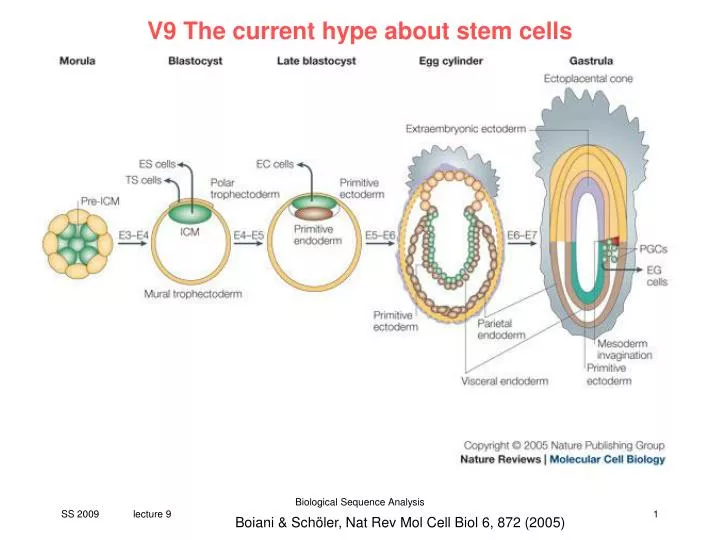
V9 The current hype about stem cells. Boiani & Schöler, Nat Rev Mol Cell Biol 6, 872 (2005). Legend of previous figure:

E N D
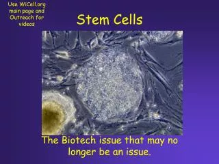
V9 The current hype about stem cells Biological Sequence Analysis Boiani & Schöler, Nat Rev Mol Cell Biol 6, 872 (2005)
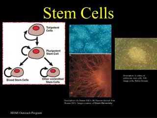
Legend of previous figure: In this figure, the pluripotent cells of the embryo are tracked in green. From left to right, the morula-stage mouse embryo (embryonic day 2.5; E2.5) holds a core of pre-ICM (inner cell mass) cells that turn into ICM cells at cavitation/blastulation (E3–E4). At this stage, embryonic stem cell (ESC) and Trophoblast Stem Cell (TSC) cell lines can be derived in vitro, and implantation occurs in vivo. The two blastocyst-derived stem-cell populations of the mouse are purported to lack the ability to interconvert. In contrast to mouse ESCs, human ESCs can be efficiently directed to differentiate into trophoblast cells, although culture conditions for establishing human TSCs have not been found so far. As the blastocyst fully expands (and undergoes implantation in vivo), the ICM delaminates giving rise to a primitive ectoderm and a primitive endoderm layer. At this stage, pluripotent cell lines that are known as embryonal carcinoma cells (ECCs) can be derived from the primitive ectoderm — whether they are distinct from ESCs has not been resolved. At E6 and subsequent stages, the experimental ability to derive ESCs, TSCs and ECCs from the mouse embryo is progressively lost, and the in vivo embryo will start gastrulating. This process involves the formation of a mesoderm layer between ectoderm and endoderm, and the formation of the primordial germ cells (PGCs). Interestingly, ESCs have been suggested to be the earliest germ cells emerging in vitro. So, the ability of ESCs to 'form' advanced germ-cell stages in vitro might not be surprising after all, and argues that ESCs (or their in vivo equivalent) give rise to specialized cell lineages by changes that direct them forwards or backwards along an existing pathway. In line with this idea it is noteworthy that pluripotent cell lines can be derived from later germ-cell stages, namely embryonic germ cells (EGCs) from PGCs, and germline stem cells (GSCs) from neonatal testis. Biological Sequence Analysis Boiani & Schöler, Nat Rev Mol Cell Biol 6, 872 (2005)
Boiani & Schöler, Nat Rev Mol Cell Biol 6, 872 (2005) Biological Sequence Analysis
Legend of previous figure: Cell-surface receptors initiate signals that are conveyed (thin black lines) to the nucleus and affect key pluripotency transcription factors such as octamer-binding transcription factor-4 (OCT4) and Nanog, and self-renewal transcription factors such as signal transducer and activator of transcription-3 (STAT3). So far, only the leukaemia inhibitory factor (LIF) receptor (LIFR)–STAT3 pathway has been defined in detail. The cytokine LIF functions by binding to LIFR at the cell surface, which causes it to heterodimerize with another transmembrane protein, glycoprotein-130 (gp130). This is followed by the activation of kinases that amplify and drive the signal to the nucleus, or that recruit other factors for docking to the activated LIFR–gp130 receptor. On binding LIF, the intracellular domains of the LIFR–gp130 heterodimer can recruit the non-receptor Janus tyrosine kinase (JAK) and the antiphosphotyrosine immunoreactive kinase (TIK) and become phosphorylated. The phosphorylated intracellular domains of the LIFR–gp130 heterodimer function as docking sites for proteins that contain Src-homology-2 (SH2) domains, which include the transcription factor STAT3. At the nuclear level, STAT3, OCT4 and Nanog cause changes in gene expression that result in (pointed arrow) or counteract (truncated line) phenotypic traits of embry-onic stem cells (ESCs). OCT4 functions in connection with SRY-related high-mobility group (HMG)-box protein-2 (SOX2) and other cofactors to determine whether target genes are activated or repressed. Aspects of this figure are speculative as, in some cases (bone morphogenic protein-4; BMP4), the surface receptors are known but the transducers are not, and in others both have yet to be identified (OCT4, Nanog). The current lack of information is highlighted by a question mark. GAB1, GRB2-associated binding protein-1; GSK3, glycogen synthase kinase-3; Id, inhibitor of differentiation, JAK, janus kinase; MEK, mitogen-activated protein kinase (MAPK) and extracellular signal regulated kinase (ERK) protein kinase; SMAD, similar to mothers against decapentaplegic homologue; SHP2, SH2-domain-containing protein tyrosine phosphate-2; WNT, wingless type protein. Biological Sequence Analysis Boiani & Schöler, Nat Rev Mol Cell Biol 6, 872 (2005)
Chronology of stem cell research • 1998 – embryonic stem cells In 1998, James Thomson (US) isolated for the first time embryonic stem cells from surplus embryos „left over“ in fertilization clinics. Since then, the research has progressed at an incredible speed. Ethics „pro“: ESC have the potential to grow replacement tissue for patients with diabetes, Parkinson or other diseases. Ethics „contra“: The technique requires destroying embryos. This has big ethical consequences. In Germany, experimentation with humans is considered problematic due to the medical experiments pursued during the Nazi time. Therefore, the above methods are forbidded by law in Germany! Researchers are looking for new ways to generate stem cells without ethical problems. Spiegel Online Biological Sequence Analysis
Chronology of stem cell research • 2006 - Induced pluripotent stem cells (iPS) The first solution was presented in August 2006 by the two Japanese Kazutoshi Takahashi and Shinya Yamanaka. Using 4 control genes, they reprogrammed cells from mouse tail into a sort of embryonic state. The product was termed induced pluripotent stem cells (iPS cells). Drawback: if used for medical treatment later, the inserted genes could enhance the risk of cancer. • 2007 – human iPS cells In 2007, similar success was managed with human skin cells. Fewer and fewer control genes are necessary to generate iPS cells. Spiegel Online Biological Sequence Analysis
Chronology of stem cell research • February 2009 – only one reprogramming gene required In February 2009, Hans Schöler presented iPS cells of mice that were reprogrammed using only a single control gene from neural stem cells (paper V9). • March 2009 – Reprogramming gene removed Begin of March 2009: 2 teams of researchers present iPS cells that do not contain additional control genes in the genome. Control genes were first inserted into the genome of human skin cells, and later removed. • March 2009 – Reprogramming gene not in genome End of March 2009: James Thomsom showed that control genes do not need to be inserted into the genome of the cells. He introduced an additional plasmid (ring genome) into the cell that was later removed. Spiegel Online Biological Sequence Analysis
Chronology of stem cell research • April 2009 – Reprogramming of mouse cells without genes Ende of April 2009: Sheng Ding (US) and others succeed to reprogram skin cells of mice into iPS without gene manipulations using proteins only. This eliminates the risk of cancer due to insertion of genes. • May 2009 – Reprogramming of human cells without genes US-korean team around Robert Lanza manages to reprogram human cells into iPS cells using proteins (TFs) only. Spiegel Online Biological Sequence Analysis
Protein Induced Pluripotent Stem Cells (PiPS) Biological Sequence Analysis
CHRONOLOGIE DER STAMMZELLFORSCHUNG Groundbreaking work demonstrated that ectopic expression of four transcription factors, Oct4, Klf4, Sox2, and c-Myc, could reprogram murine somatic cells to induced pluripotent stem cells (iPSCs), and human iPSCs were subsequently generated using similar genetic manipulation. To address the safety issues arose from harboring integrated exogenous sequences in the target cell genome, a number of modified genetic methods have been developed and produced iPSCs with potentially reduced risks. However, all of the methods developed to date still involve the use of genetic materials and thus the potential for unexpected genetic modifications by the exogenous sequences in the target cells. Zhou, Cell Stem Cell 4, 381 (2009) Biological Sequence Analysis
CHRONOLOGIE DER STAMMZELLFORSCHUNG To generate recombinant proteins that can penetrate across the plasma membrane of somatic cells, we designed and fused a poly-arginine (i.e., 11R) protein transduction domain to the C terminus of four reprogramming factors: Oct4, Sox2, Klf4, and c-Myc ... We found that the purified 11R-tagged recombinant transcription factors readily entered cells at concentrations of 0.5 8 g/ml within 6 hr and could translocate into nucleus. In addition, we found that the transduced proteins appeared to be stable inside cells for up to 48 hr. Zhou, Cell Stem Cell 4, 381 (2009) Biological Sequence Analysis
CHRONOLOGIE DER STAMMZELLFORSCHUNG We then employed this simple protein transduction protocol to reprogram OG2/Oct4-GFP reporter MEF cells. Because reprogramming through the iPSC mechanism/process typically requires sustained activity of reprogramming proteins for 7 - 10 days, we devised a strategy that involved treating the cells in four cycles. Zhou, Cell Stem Cell 4, 381 (2009) Biological Sequence Analysis
CHRONOLOGIE DER STAMMZELLFORSCHUNG Zhou, Cell Stem Cell 4, 381 (2009) Biological Sequence Analysis