Download
1 / 22
230 likes | 243 Views
بسم الله الرحمن الرحيم Practical medical mycology Lab 2 Specimen collection and transportation. The most important steps for the isolation of fungus are: 1- Proper collection of specimens. 2- Rapid transport to the lab. 3- Prompt and correct processing of the specimen.

E N D
بسم الله الرحمن الرحيم Practical medical mycology Lab 2 Specimen collection and transportation
The most important steps for the isolation of fungus are: 1- Proper collection of specimens. 2- Rapid transport to the lab. 3- Prompt and correct processing of the specimen. 4- Inoculation onto appropriate media. 5- Incubation at suitable temperatures.
General considerations •All specimens should be collected aseptically and placed in sterile containers. •Swabs are not encouraged, but specimens from the ear canal, nasopharynx, throat, vagina and cervix are not easily collected by any other means. •Many fungal infections are clinically similar to mycobacterial infections and often the same specimen is cultured for both fungi and AFB. •If a specific fungal agent is suspected, note on the request. Include travel and/or exposure history. 3 Dr. Nabil El Aila Med. Mycology
Transport of Specimens • Specimens should be transported in a sterile, humidified, leak-proof container. • Dermatologic specimens, however, should be transported in a dry container. • Specimens should be processed and inoculated to primary isolation media as soon as possible after collection, ideally within a few hours.
Transport of Specimens • If transport is delayed, store specimens under refrigeration at 4°C with the following exceptions: • - Hold blood, bone marrow and CSF at body temperature at 30-37 °C. • - Hold dermatological specimens at room temperature. • - Be aware that viability may decrease with prolonged storage.
Collecting specimens for the investigation of fungal infections •Fungal infections of the skin, nails and hair are caused by dermatophytes. •In many cases, diagnosis of infection is made by clinical appearance alone, but laboratory examination of skin scrapings, nail cuttings and hair can be requested if the diagnosis is uncertain.
Fungal infections are caused by dermatophytes •Fungal infections of the skin, nails and hair are caused by dermatophytes, which require keratin for nutrition. •The estimated lifetime risk of acquiring a superficial fungal infection is between 10 – 20%, although these are rarely, if ever, invasive.
Organisms involved in fungal infections •Superficial fungal infections may be caused by one of over forty different species of dermatophytes, belonging to the following three genera; •Trichophyton spp – found in hair, nails and skin, transmitted by soil, animals or humans •Microsporum spp – common cause of scalp ringworm in children, usually transmitted by animals •Epidermophyton spp – most commonly affects the groin, transmitted from person to person.
When do fungal specimens need to be collected? •Minor localized infections can be treated topically without the need for fungal testing. •Specimens should be sent to confirm disease when the infection is chronic, severe or when considering systemic therapy.
How to collect a suitable specimens •In most cases, collection of fungal specimens is performed at the laboratory. If this is not possible, or if the clinician wishes to collect the sample themselves, the following guidance may be helpful. •Ensure that the patient has not used anti-fungal medications for the previous three days. •If collection of the specimen is proving difficult, then consider asking the patient to do it themselves, under supervision. •Patients are often more aggressive at getting a good sample than a collector who is trying to be gentle.
Laboratory fungal testing is also justifiable in the following circumstances: - To confirm fungal infection before starting on oral treatment, e.g. if the patient has been treating the lesion with topical steroids or a fungal infection involving the hair, palms of the hands or soles of the feet. - To determine the species of fungus to allow targeted oral treatment - On epidemiological grounds, e.g. people in contact with an animal in cases of animal ringworm.
Mycology Specimen Collection Protocol A) Specimen Collection, transportation and storage • Hair: • No cleaning of scalp is needed. • Select infected areas and with forceps, epilate at least 10 hairs. •For hairs broken off at the scalp level, use a scalpel or a blade knife. •Place hairs between two clean glass slides or in a clean envelope labeled with the patient's data.
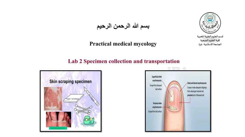
2. Skin: • Wipe lesions and interspaces between the toes with alcohol sponge or sterile water. • Scrape the entire lesion(s) and both sides of interspaces with a sterile scalpel. • Place scrapings between two clean glass slides or place in a clean envelope labeled with the patient's data.
3. Nails •Clean nail with 70% alcohol. •Dorsal plate - Scrape outer surface and discard; scrape the deeper portion. •Remove a portion of debris from under the nail with a scalpel. •Collect whole nail or nail clippings. •Place all material in a clean envelope labeled with the patient's data.
4.Cerebrospinal fluid • Cerebrospinal fluid is collected aseptically by needle aspiration and should be sent to the laboratory in a sterile container as quickly as possible . • The fluid should be concentrated by centrifugation for 15 minutes at 1000 x g, and a minimum of 0.5 ml of sediment should be inoculated onto the surface of media, but the small volumes of CSF not suitable for centrifugation may be dropped directly on media surface. CSF specimens stored at 25-30°C.
5.Blood • Ten milliliters of blood should be collected in EDTA tube after good skin antisepsis and stored at 25-30°c. 6. Bone marrow • Bone marrow aspirates and biopsies are collected after good skin antisepsis and are commonly submitted to the Laboratory in a sterile syringe or tube containing EDTA. • The collected specimen should be transported to the Laboratory as soon as possible; however, it may be refrigerated at( 2-8 c) for no longer than 12 hours, if immediate culturing is impossible.
7. Urine • At least 10ml of first morning specimen, suprapubic aspirates, midstream or catheterized specimen are collected. • Specimens should be cultured promptly since bacteria and yeasts replicate rapidly in specimens kept at room temperature. • If specimens cannot be cultured soon after receipt, they should be refrigerated at 4°C for no longer than 12 to 15 hours.
8. Vaginal secretions • Vaginal and cervical specimens collected by a physician are usually submitted on a swab. • Transport to the laboratory should be rapid; however overnight refrigeration before culturing is satisfactory. Specimens should not be stored at room temperature.
9. Respiratory specimens • Specimens from the nose, nasopharynx , and mouth are usually submitted on a sterile swab. • A first morning expectorated sputum specimen should be collected in a sterile wide-mouth bottle or sputum cup. • Specimens should be transported to the laboratory as soon as possible to ensure maximum recovery of fungi. • If culturing is delayed, specimens may be refrigerated at 2-8°C.
10. Tissues/biopsies • Tissue should be divided aseptically by the surgeon in the operating room, all biopsy tissues should be placed in a sterile container containing a small amount of saline without a preservative. • When an abscess is drained, a portion of the abscess wall should be submitted for culture. The surgical pathologist will visually examine specimens prior to submission to the microbiology lab. • Surgical specimens should be transported to the surgical pathology laboratory as soon as possible after collection. If immediate culturing is impossible, specimens should be stored at 4°C for no longer than 8-10 hours.